Scientists use fat-based ‘bubbles’ to sneak ALS treatment into the brain
Approach bypasses blood-brain barrier to deliver GM1 to affected nerve cells
Written by |
- Fat-based 'bubbles' (talineuren) deliver GM1 past the blood-brain barrier for ALS treatment.
- In mouse models, talineuren improved motor function and extended survival by repairing motor neurons.
- These promising findings pave the way for testing in future human clinical trials.


Scientists have developed a way to deliver a nerve-protecting substance past the brain’s natural defenses using fat-based ‘bubbles,’ a strategy that significantly extended survival and preserved movement in mouse models of amyotrophic lateral sclerosis (ALS).
This approach, called talineuren, successfully delivered the protective molecule GM1 directly to damaged motor neurons, offering a potential path toward a disease-modifying treatment for patients.
The study, led by Smita Saxena, PhD, at the University of Missouri School of Medicine, found that packaging the protective molecule GM1 into specialized vesicles enabled it to cross the blood-brain barrier and repair damaged nerve cells. These findings pave the way for future human clinical trials at the Roy Blunt NextGen Precision Health building, where researchers aim to translate these laboratory successes into patient care.
“The NextGen Precision Health building is the perfect place for this research,” Saxena said in a university news story. “By having research and clinical space under the same roof, we can speed up the process for translating foundational research into human clinic trials to ultimately help improve quality of life for Missourians and people worldwide.”
The study, “Engineered GM1 Intersects Between Mitochondrial and Synaptic Pathways to Ameliorate ALS Pathology,” was published in Advanced Science.

Cellular stress and the breakdown of motor neurons
ALS damages motor neurons, the nerve cells that control voluntary movements. With the loss of these nerve cells, the brain is no longer able to control muscles. As a result, patients gradually lose the ability to perform everyday tasks, such as walking and reaching for items.
Earlier work by Saxena’s research team showed that motor neurons in ALS become highly sensitive to stress in the endoplasmic reticulum, a structure inside cells that helps proteins fold into their correct shape. When the endoplasmic reticulum is stressed, proteins misfold and build up as toxic aggregates that damage nerve cells. This stress also affects mitochondria, the structures in cells that produce energy.
“Our research team’s previous work showed that neurons in those with ALS are very sensitive to endoplasmic reticulum stress, which limits the mitochondria’s ability to produce energy,” said Saxena, who is also an investigator at NextGen Precision Health. “This, in turn, limits the ability of neurons to send messages across synapses [the junctions where nerve cells communicate] when we want to move our muscles.”
Treating ALS has been difficult partly because of the blood-brain barrier, a tightly controlled layer of cells that protects the brain from toxic substances in the bloodstream. However, it also blocks the entry of treatments.
GM1 is a naturally occurring fat-like substance that helps nerve cells survive, communicate, and repair upon damage. The compound has shown promise in ALS mouse models, but its limited ability to cross the blood-brain barrier has led to mixed results in clinical studies.
To overcome this, Saxena partnered with Innomedica to use talineuren, a nanoliposome (a fat-based vesicle) that contains GM1 in its membrane and can cross the blood-brain barrier to deliver the compound to the affected nerve cells.
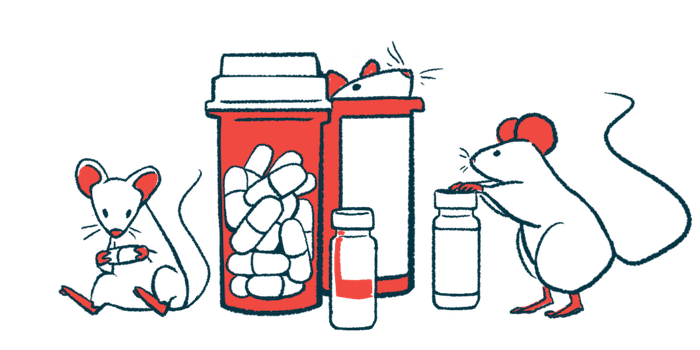
In mouse models carrying ALS-causing mutations in the C9orf72 and SOD1 genes, talineuren improved motor performance. Mice given talineuren maintained stronger grip strength and performed better in coordination on movement tests than untreated animals. These functional gains suggest the treatment helped preserve motor neurons and delay motor deficits.
In the C9orf72 model, the treatment also significantly extended lifespan in both male and female mice, regardless of disease progression rate. Median survival increased by more than 121 days in males, 115 days in slowly progressing females, and 35 days in rapidly progressing female mice.
At the cellular level, the treatment helped correct several biological processes disrupted in ALS. For example, talineuren reduced stress in the endoplasmic reticulum, stabilized calcium handling in mitochondria, and improved energy metabolism. The treatment also prevented the formation of toxic protein clumps, restoring the cell’s ability to maintain a healthy protein balance.
Importantly, the treatment was well tolerated, with no major safety concerns reported in the study, positioning talineuren “as a promising therapeutic candidate,” the researchers wrote.
The treatment has been granted orphan drug designation for ALS in the U.S. and Europe, a status that provides a range of incentives to develop treatments for rare diseases. According to the researchers, this sets “the stage for future human clinical trials.”