MRI Could Serve as Useful Biomarker to Assess ALS Progression, Study Suggests

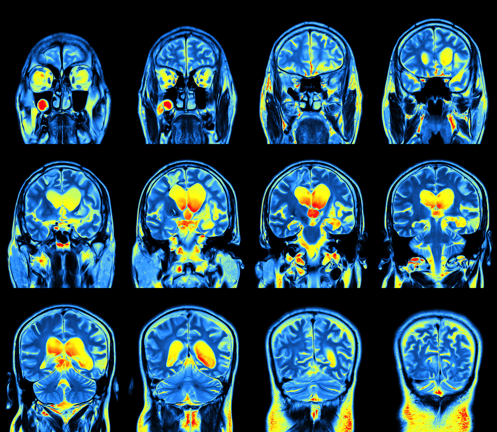
Magnetic resonance imaging (MRI) can detect short-term changes in the brain caused by amyotrophic lateral sclerosis (ALS), according to a study in Neuroimage: Clinical.
This makes the MRI a good neuroimaging marker to follow the progression of ALS by helping doctors design better clinical trials and assess how well patients respond to a certain treatment.
For the study, “Longitudinal evaluation of cerebral and spinal cord damage in Amyotrophic Lateral Sclerosis,” a team of researchers led by Dr. Marcondes Cavalcante França Jr. of Brazil’s University of Campinas assessed 27 ALS patients and 27 healthy controls clinically and using MRI. They repeated the assessments twice, eight months apart.
The team quantified the severity of the disease at both timepoints using a test called ALS Functional Rating Score-Revised (ALSFRS-R), which measures daily living activities and global function of ALS patients. They also analyzed MRI-derived measures such as the thickness of the cerebral cortex (the brain’s outer layer) and the neck spinal cord.
The team, whose research was funded by the São Paulo State Research Foundation, that although the cerebral cortex showed no progressive thinning in the cortex of the brain, people with ALS did show a significant reduction in the volume of their brainstem after eight months.
The researchers also saw an increase in diffusivity, or the ability of water molecules to pass through a region of the brain called the corpus callosum. This relates to the loss of cerebral white matter integrity, a hallmark of ALS. However, these changes did not correlate with changes in the disease’s severity or duration.
The reduction in the size of the neck spinal cord was the only MRI parameter that correlated with a change in ALSFRS-R, or disease severity, leading researchers to conclude that measuring the size of the spinal cord in the neck is a promising neuroimaging marker to assess ALS progression.
They urged further research in a larger group of patients to assess the relationship between spinal cord damage and functional impairments in ALS.


