Retinal Layer Thinning May Help Track Disease Progression in ALS Patients

Amyotrophic lateral sclerosis (ALS) patients without any type of eye disease have thinner retinal layers, a new study shows.
The research, titled “Retinal thinning in amyotrophic lateral sclerosis patients without ophthalmic disease” was published in the journal PLOS ONE.
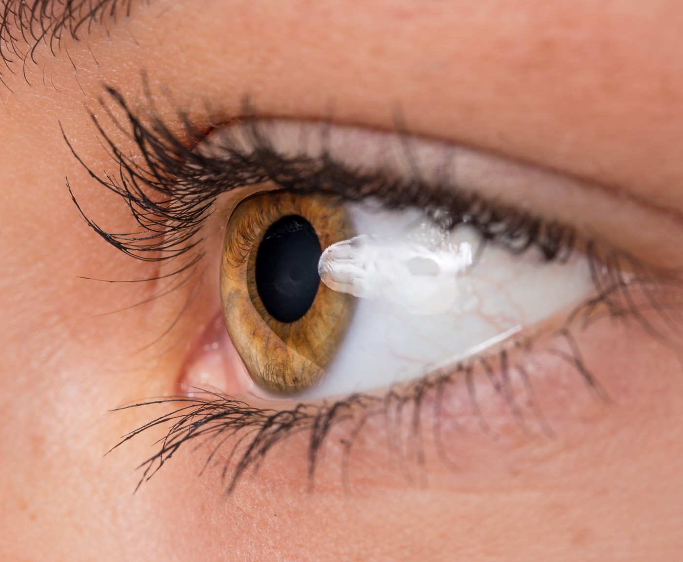
The retina is the innermost coat at the back of the eyeball that contains light-sensitive cells. These convey “light information” to the optic nerve, which then transmits the message to the brain, where an image is formed.
ALS is a disease characterized by motor neuron degeneration. Studies have shown there are three causative genes associated with both ALS and glaucoma (an eye condition that if left untreated may cause blindness). But whether ALS patients have neurodegeneration in their retinas that is not associated with ocular diseases is an unanswered question.
To investigate this hypothesis, researchers developed an observational, comparative, cross-sectional study to determine if ALS patients had differences in retinal neurons.
In the study, 21 ALS patients were assessed with spectral domain optical coherence tomograph (SD-OCT), a technique that allows measurement of retinal nerve fiber layer (RNFL) thickness.
Results showed that ALS patients had significantly thinner retinal layers compared to values in the SD-OCT normative database (88.95 microns vs. 95.81 microns). Eye diseases that could have an effect on retinal thickness were excluded through detailed history and an ophthalmic examination.
Therefore, findings support the team’s initial theory that retinal thinning most likely is associated with ALS-related neurodegeneration.
Interestingly, the RNFL thickness values were not correlated to ALS disease severity, intraocular pressure, or visual acuity.
This discovery is important in a medical diagnosis kind of way, as clinicians can consider using SD-OCT in an outpatient clinic, as a rapid, non-invasive tool to track disease progression in ALS patients.
However, there is still a need to deepen the understanding of how the retinal thinning correlates to ALS disease progression.
“Further clarification of the temporal relationship between motor neuron degeneration and retinal neurodegeneration in ALS patients or animal models will aid in the use of the retina as a tool to monitor ALS disease progression and assess responses to potential therapies,” the team concluded.


