New Rat Model Enables Better Study of Swallowing Issues in ALS, Researchers Say

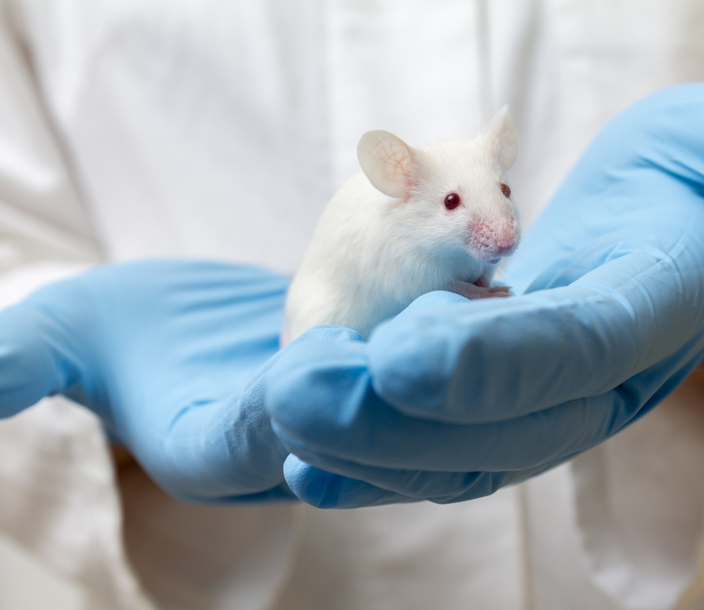
A new, more targeted rat model of swallowing problems may lead to better understanding of ways to preserve and even restore tongue function in patients with amoytrophic lateral sclerosis (ALS), according to a study.
The study, “Hypoglossal Motor Neuron Death Via Intralingual CTB-saporin (CTB-SAP) Injections Mimic Aspects of Amyotrophic Lateral Sclerosis (ALS) Related to Dysphagia,” was published in the journal Neuroscience.
Swallowing impairment, or dysphagia, may be an early sign of ALS, resulting from degeneration of motor neurons — cells that control muscle contraction — in the hypoglossal nucleus in the brainstem. Nerve fibers arising from this nucleus innervate the tongue. As a result, progressive motor neuron death leads to tongue weakness, an essential muscle for swallowing, breathing, and speaking.
Studying dysphagia in ALS models has been challenging, as the amount and rate at which hypoglossal motor neuron death occurs cannot be controlled, and because degeneration is not limited to this nucleus.
Aiming to address this gap, a team from the University of Missouri School of Medicine and the University of Missouri College of Veterinary Medicine developed a model in rats that involves into-the-tongue injections of a tracer protein called cholera toxin B conjugated to saporin (CTB–SAP), a toxin and so-called ribosome-inactivating protein that inhibits protein synthesis and growth, leading to neuronal death.
“We created a rodent model that allows us to study only the tongue,” Teresa Lever, PhD, a study co-author and a professor of otolaryngology at the MU School of Medicine, said in a press release. “By targeting a muscle we know is affected by ALS, we hope to better understand how we might eventually slow ALS’ ability to compromise multiple upper airway functions.”
Upon CTB–SAP injection into the tongue’s genioglossus muscle, the rats showed statistically significant deficits in lick and swallow rates, hypoglossal motor output, and hypoglossal motor neuron survival compared with controls.
The rats also exhibited compensatory behavioral changes — such as placing a forepaw on the edge of the bowl when drinking — similar to the well-known SOD1G93A ALS model. In addition, injected rats held their head in a more vertical position while drinking, which resembles the “dropped head syndrome” seen in ALS patients.
With this approach, the investigators were able to restrict disease-related alterations to hypoglossal motor neuron death only, without the diverse complications observed in ALS models
“This novel, inducible model of hypoglossal motor neuron death mimics the dysphagia phenotype that is observed in ALS rodent models, and will allow us to study strategies to preserve swallowing function,” the scientists wrote. “Perhaps most importantly, the rate and degree of neuronal loss was consistently controlled in our model, which could be advantageous to test new therapies.”
“We have developed a unique rodent model that has reproducible hypoglossal motor neuron death and only tongue dysfunction,” said Nicole Nichols, PhD, the study’s senior author and a professor of biomedical sciences at the MU College of Veterinary Medicine.
Nichols further said that the model enables them to look at whether the surviving motor neurons adapt to the loss of the others, adding, “What we want to do next is try to make the surviving neurons work a little harder to show that we can improve tongue function both in the short and long term.”


