Pooling Imaging Data May Make Multi-center Clinical Trials Easier


Imaging data of brain inflammation in people with amyotrophic lateral sclerosis (ALS) that is obtained using different tracers and imaging equipment can be combined using a technique called the “pseudo reference region approach.”
This finding may make it easier to conduct multi-center studies about ALS, according to a recent study.
The study, “Moving Toward Multicenter Therapeutic Trials in Amyotrophic Lateral Sclerosis: Feasibility of Data Pooling Using Different Translocator Protein PET Radioligands,” was published in the Journal of Nuclear Medicine.
Brain inflammation is one of the hallmark biological features of ALS. As such, measuring the extent of inflammation in the brain is thought to be a promising approach for assessing disease severity and responses to treatment. Such measurements are particularly important in the context of clinical trials, since it is necessary to have objective measurements to determine whether an investigational treatment has a real effect.
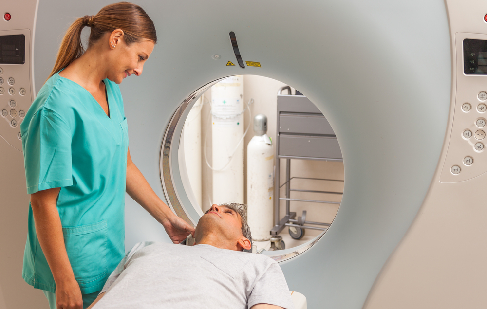
Brain inflammation can be detected using imaging technology in combination with certain radioactive tracer molecules that are taken up by activated inflammatory cells in the brain, namely those producing the translocator protein (TSPO).
A challenge in doing large studies on ALS is that different institutions often have different available equipment and setups, and it is typically difficult to accurately make comparisons among data that is collected through different techniques.
Two promising candidate TSPO tracers for ALS and other neuroinflammatory diseases, 11C-PBR28 and 18F-DPA714, are in development in the U.S. and Europe, respectively.
Now, researchers have shown that it is possible to pool data obtained with 11C-PBR28 and 18F-DPA714 by using the pseudo reference technique.
The gold standard for imaging with TSPO tracers involves full dynamic modeling using arterial blood sampling, which measures levels of tracers in the blood in order to make determinations about where in the body the tracer is accumulating (indicating inflammation). The pseudo reference technique essentially uses part of the brain image itself, instead of taking a blood sample, in order to make these determinations.
“This technique reduces patient burden, scan time, and the discomfort of undergoing arterial line placement, compared with the dynamic modeling technique,” the researchers wrote.
“Furthermore,” they added, “this technique might enable multicenter pooling of different TSPO radioligands,” because using a ratio based entirely on imaging data removes possible complicating variables, such as differences between cameras or levels of tracers.
The researchers analyzed data from seven ALS patients and eight healthy volunteers who underwent imaging with 18F-DPA714 in Belgium, and from seven ALS patients and seven healthy volunteers who were imaged with 11C-PBR28 in the U.S.
“This study was an initial effort to combine and compare data acquired from 2 separate cohorts of ALS participants with separate tracers and 2 different scanners, using a fully automated analysis pipeline and pseudo reference region analysis technique. To the best of our knowledge, there have been no prior direct comparison studies of 18F-DPA714 and 11C-PBR28 PET scans,” they wrote.
Compared to the healthy controls, in ALS patients, there was greater uptake of both markers in parts of the brain that control movement, a finding that is consistent with increased brain inflammation in ALS patients.
Statistical analyses demonstrated that findings with each of the two tracers were comparable, meaning the data feasibly could be pooled while still retaining accuracy.
“We have shown the ability to pool together and analyze brain neuroinflammation PET imaging data acquired at multiple institutions with varying scanner capabilities, using state-of-the-art analytical tools,” co-author Suma Babu, assistant professor of neurology at Harvard Medical School, said in a press release.
“This approach could reduce time and travel burden for patients, allowing them to participate in novel biomarker research while remaining close to home,” Babu added. “From a scientific study conduct standpoint, this approach retains scientific rigor, increases statistical power, reduces trial durations and reduces risks of attrition.”
Being able to pool data collected at different institutions using different techniques could make it easier to conduct multi-center clinical trials, the researchers said.
“In rare diseases such as ALS, only a limited pool of participants is available to participate in imaging studies. Therefore, conducting collaborative research across various sites and bringing in data to a common analysis pool is valuable to accelerate imaging biomarker development,” said co-author Donatienne Van Weehaeghe, MD, PhD, of University Hospital Leuven in Belgium.
The researchers noted several limitations to this study, including the small sample size, and the fact that each person was imaged with only one of the tracers, making it impossible to do direct comparisons. They are planning more studies with more participants across a wider range of disease severities.