
Damage to Brain Areas Crucial to Thinking, Not Motor Neurons, Seen as Root of ALS Cognitive Problems
Written by |

Amyotrophic lateral sclerosis (ALS) patients with cognitive difficulties show brain damage in areas important to thinking, a finding that may explain why such patients deteriorate more rapidly.
The study, “Structural explanation of poor prognosis of amyotrophic lateral sclerosis in the non-demented state,” published in the European Journal of Neurology, also supports the theory that ALS disease processes can start either in the brain’s cortex or in the spinal cord.
Studies showing that ALS is likely a disease involving several brain regions, in addition to motor neurons, have been steadily emerging in recent years. ALS patients can have both cognitive and behavioral problems, and the condition frequently coexists with frontotemporal dementia.
Researchers at Hanyang University in Korea figured that neuropsychological differences among patients may be mirrored by damage to corresponding brain areas.
The research team recruited 47 patients with sporadic ALS who did not have dementia, and 28 healthy controls. Controls were matched with patients according to age, to improve the validity of the analysis. After testing the patients’ neurologic and cognitive abilities, the team divided them into three groups: patients with cognitive impairment, patients with behavioral problems, and those with “pure” ALS (motor or movement difficulties).
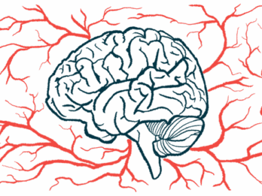
All participants had their brains scanned with a magnetic resonance imaging (MRI) technique that can assess the volume of particular brain regions.
First, the team compared images between the different patient groups and controls to identify affected brain regions. It soon became apparent that patients with cognitive problems showed brain tissue loss in areas that were not affected in the other patient groups.
Specifically, only patients with cognitive problems had gray matter tissue loss in the cerebellum and an area of the cortex involved in cognitive processing. Researchers also found loss of white matter in regions processing visual signals, and in nerve pathways connecting the brain and spinal cord. In contrast, patients with behavioral problems or “pure” ALS had no significant brain volume loss compared to healthy controls.
Correlating brain imaging data with neuropsychological assessments, the team also found that patients with cognitive problems had volume loss in brain areas involved in fine-tuning and coordinating movement, including important regions connecting different areas.


Leave a comment
Fill in the required fields to post. Your email address will not be published.