
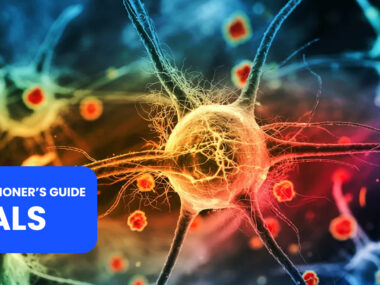
Astrocytes and Microglia in ALS: Friends Turn to Foes
Written by |
A report in the journal Experimental Neurobiology reviewed the role of astrocytes (cells that support and protect neurons) and microglia (immune system cells in the brain) in amyotrophic lateral sclerosis (ALS).
The authors, from the Veterans Affairs Boston Healthcare System; the Department of Neurology at Boston University School of Medicine; and the Center for Neuromedicine, Brain Science Institute, Korea Institute of Science and Technology in Seoul, sought to understand those particular mechanisms behind ALS and the disease’s progression.
For the study, “Astrocytes And Microglia As Non-Cell Autonomous Players In The Pathogenesis Of ALS,” the authors reviewed reports revealing several aspects about the involvement of non-neuronal cells in the development of ALS.
Scientists had long believed that neuronal damage associated with ALS resulted from improper function and regulation in neurons themselves — not related to dysfunction of other brain cells.
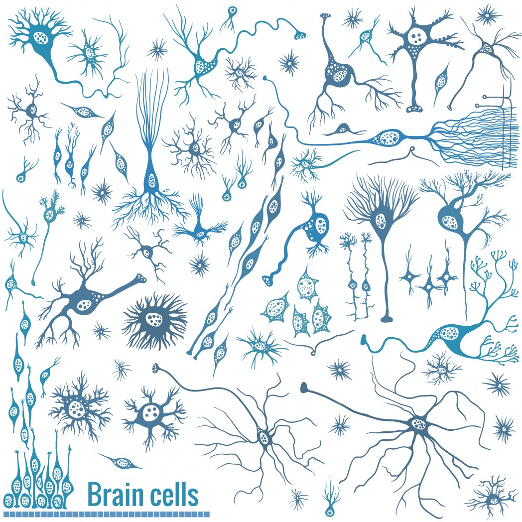
Changes in other cells present in the brain, such as astrocytes and microglia, accompany neuronal anomalies that contribute to ALS and play a crucial role in the damage and death of motor neurons. Although their activity is fundamental for neuronal health and maintenance under normal conditions, in ALS the cells turn from friends into foes.
Regarding the role of astrocytes, the authors reviewed several studies showing that inflammatory proteins (cytokines) produced by these cells can enhance disease progression and are critical to the development of ALS.
EAAT2 is another astrocytic protein that plays a role in the disease. Under normal conditions, EAAT2 transports glutamate (a molecule that stimulates neuronal activity in the brain) back into astrocytes when communication between neurons is over, allowing neurons to rest until a new signal for activation comes in.
But in ALS, the expression of the EAAT2 gene is decreased and glutamate accumulates in excessive levels around neurons, leading to their abnormal activation and damage. Regulation of the astrocytes’ EAAT2 then contributes to motor neuronal survival and death in ALS.
For the role of microglia, the authors explained that its function is necessary for surveilling motor neuron conditions and for restoring tissue injury in response to acute and reversible stress.
“Microglia are beneficial before the threshold limit reached. However, constitutive activation of microglia by a chronic and irreversible stress such as ALS stress may [cause these cells] to be toxic to motor neurons: microglia are disadvantageous after they become fully activated,” the authors wrote.
In the review, the authors discuss studies that raised questions about whether different stages of microglia activation are involved in different modes of action (protection versus damage of motor neurons) through yet unidentified mechanisms.
More studies are necessary “to uncover the precise action mechanism behind the obscure role of microglia in ALS,” the authors wrote.
Overall, regarding ALS, cells that in normal conditions protect neurons act differently. They undergo a series of molecular and cellular changes that affect the health and activity of motor neurons, leading to irreversible damage and loss. Reactive astrocytes and microglia trigger neuroinflammation, which accelerates the progression of the disease by increasing ongoing neuronal injury.
Findings concerning the contribution of astrocytes and microglia “suggests that future therapeutic strategy for the treatment of ALS should be aimed at specific interception of pro-oxidant and pro-death signals in a cell-type specific manner,” the authors wrote.

Leave a comment
Fill in the required fields to post. Your email address will not be published.