
Mouse Study Changes Perspective on Brain Immune Cells’ Role in ALS Progression
Written by |
Some immune cells in the brain can protect it from the damaging effects of faulty TDP-43 protein, preventing the progression of amyotrophic lateral sclerosis in mice, University of Pennsylvania researchers report.
The findings challenged a long-held assumption that the immune cells play a role in damaging nerve cells, the team said.
Their study, published at Nature Neuroscience, was titled “Microglia-mediated recovery from ALS-relevant motor neuron degeneration in a mouse model of TDP-43 proteinopathy.”
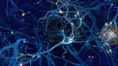
Until now, scientists had considered the activation of immune cells in the brain known as microglia to be a hallmark of ALS. But a Perelman School of Medicine team at the University of Pennsylvania discovered that a subset of microglia can actually protect nerve cells.
“The prevailing view in the field has been that immune system inflammation contributes to the death of neurons in ALS, but this study shows the opposite — that microglia are actually critical for neuronal [nerve cell] survival,”Dr. Virginia M-Y. Lee, director of the university’s Center for Neurodegenerative Disease Research, said in a news release. She was senior author of the study.
The team engineered a mice model of ALS to mimic the TDP-43-driven human disease. TDP-43 accumulates in the nerve cells that control movement, contributing to ALS’s progression.
An analysis of the mice’s brains and spinal cords at different stages of their disease indicated that rising levels of TDP-43 had only a mild effect on the immune cells there. The protein’s accumulation did not change the number of immune cells or their activity, even when there was considerable nerve cell death.
Interestingly, when the team used a chemical to prevent further accumulation of TDP-43 at early stages of the disease, the number of microglia cells increased by 70 percent. This was not due to infiltration of new immune cells but from the proliferation of cells already there. Another finding was that microglia cells changed their shape and their genetic-activity profile.
The difference in response based on the presence or absence of TDP-43 surprised the team. “This is still a mystery, but one that we’d very much like to figure out in future studies,” said Dr. Krista J. Spiller, the study’s lead author.
An important finding was that the immune cells targeted the nerve cells with clusters of TDP-43 protein. This neuroprotective activity led to a reversal of the mice’s movement symptoms, with some regaining their ability to walk. But when the researchers prevented the microglia cells from acting, the mice were unable to regain full muscle function.
“The way reactive microglia protect neurons [nerve cells] points us towards new ideas for ALS therapies,” Spiller said. “For example, we want to know if we can encourage the expansion of microglia in early-stage ALS patients to save their motor neurons [movement nerve cells].”
While previous studies had suggested that targeting microglia might be a way to slow the progression of ALS, the new finding support an opposite strategy, the researchers said. In other words, a strategy of “findings ways to encourage appropriate microglia-mediated inflammation during the onset and progression of ALS to clear pathological TDP-43 proteins and boost axonal [movement nerve cell] regeneration.”
Improved understanding of the underlying mechanism of microglia’s neuroprotective role “may provide new therapeutic strategies for patients even in late disease stages,” the team added.


Charlie
"It's bad..."
"No...It's good..."
"Just a minute...."
"Er... wait a moment..."
"Mmmm...er... hold on....."