
Transplanted Human Bone Marrow Cells Hold Promise as ALS Therapy, Mouse Study Shows
Written by |
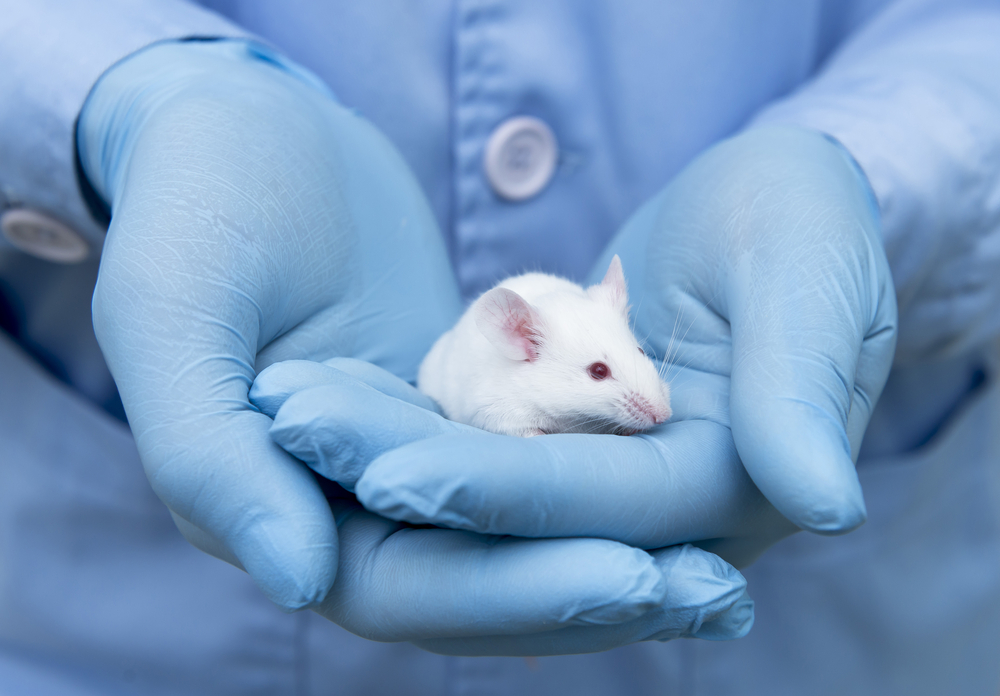
Transplanting certain human bone marrow cells into mice with amyotrophic lateral sclerosis (ALS) improved their motor function and preserved motor neurons by repairing the barrier protecting the spinal cord.
The study with that finding, “Human Bone Marrow Endothelial Progenitor Cell Transplantation into Symptomatic ALS Mice Delays Disease Progression and Increases Motor Neuron Survival by Repairing Blood-Spinal Cord Barrier,” was published in the journal Scientific Reports.
The blood-brain and blood-spinal cord barriers (BSCB) are composed of endothelial cells, pericytes, located along the walls of tiny blood vessels called capillaries, and processes derived from astrocytes, a cell type also involved in the response to injury.
Increasing evidence has linked impairments in each of these components to ALS, both in patients and in animal models. Disrupted barriers allow the entry of immune cells and other potentially harmful substances from the blood circulation, which may aggravate degeneration of motor neurons — specialized cells that control muscle contraction — a hallmark of ALS.
In mice with ALS, the team at University of South Florida in Tampa previously showed that human bone marrow cells containing the CD34 marker (hBM34+) may be a therapeutic stray for ALS, as they delayed disease progression, preserved motor neuron survival, lessened a marker of reaction to injury, and maintained barrier integrity. The transplanted cells differentiated into endothelial cells — those that line blood vessels — and nested in capillaries of the spinal cord.
Join our ALS forums: an online community especially for patients with Amyotrophic Lateral Sclerosis.
A subsequent study in the same mouse model, G93A, revealed that a high dose of transplanted cells restored the fine structure of capillaries and stabilized their density in the spinal cord, while also improving myelin integrity. (Myelin is the protective layer that insulates the nerves.)
As noted in a press release, although these findings support the use of such cells to repair the BSCB and improve ALS-related alterations, the most significant effect of hBM34+ cells on motor function was determined at four weeks after transplant. Also, a substantial damage in spinal cord capillaries was detected even after high-dose treatment.
This made the team test whether human endothelial progenitor cells (EPCs) — bone marrow-derived, but more similar to endothelial cells than undifferentiated stem cells — would provide superior BSCB restoration in G93A mice.
At two to three weeks after transplant via intravenous infusion, mice given EPCs from adult donors showed significantly higher body weight, improved motor function and slowed disease progression than controls with ALS, as assessed by extension reflex scores, grip strength, and motor coordination.
At four weeks, these mice also demonstrated BSCB repair associated with widespread attachment of EPCs to capillaries in the cervical and lumbar spinal cord and in the brain’s motor cortex and brainstem — two regions known for motor neuron degeneration.
Other improvements in the cervical and lumbar spinal cord included restored capillary and pericytes’ structure, normalized astrocytic processes, less leakage into the spinal cord, and extended survival of spinal cord motor neurons. This was reflected by signs of degeneration in only a small subset of motor neurons and by higher number of these nerve cells than in controls with ALS.
Cautioning that further studies are needed, the team commented that “from a translational viewpoint, the initiation of cell treatment at the symptomatic disease stage offered robust restoration of BSCB integrity and shows promise as a future clinical therapy for ALS.”


Dave Reckonin
Looks Good.
A Big-Pharma spokesperson commented 'Oh Noooooooooooooo"
Geo
Great news - introduce our own Bone Marrow Cells to correct the spinal cord defects caused by disease and injury.
How do we trsnslate this research quickly to realize some hope for so many suffering each day. My God help us to move faster.
Dave Reckonin
So Sorry. God has proved he is disinterested.
Fran
I can understand arguments for atheism. But your interest in taking away the comfort others choose to take from their faith seems cruel. If you may be suffering pain that's inspiring your attacks, I'm sorry for you.
Dave Reckonin
I have no interest and certainly no time available for taking anything away from people who choose to believe.
I doubt I could do even if I wanted to. Most God-botherers cannot see farther than their own nose -ends that the entity they believe in, without logic, rationality or reason, has not responded to millions of prayers or supplications for easement of ALS.
I do not attack anyone, but I am roundly attacked and ridiculed by the army of God-Botherers
Anyhow, it seems very clear that, should he exist, he is disinterested in the plight of pALS.
I do not require your sorrow either. You sound profoundly patronizing by saying this and I suspect it comes, along with your anger, from your suspicion that the God you believe in so strongly has abandoned you or does not exist.
He cannot be 'all-loving' and yet desert you in the face of ALS.
Is it so difficult for you to believe in the logic of the five Natural Sciences, particularly Biology, where nature has decreed a defective gene pattern that kills pALS in such a brutal UnGodly way?
This is not difficult to understand if you disallow dogma from clouding your thinking.
Darned if I know how comfort arises from Religion for pALS going through this torture. It seems a logical reason to avoid its mantras and illogicalities.
jan berkhey
no, god is a healer, sickness is not his fault, he healed us 2000 years ago when jesus died on the cross
Dave Reckonin
God, a 'healer'? A 'healer'?
Do you realize just how ridiculous your claim sounds?
God heals nothing. Science tries and tries and tries to heal things.
God destroyed Sodom & Gomorrah, yet he did not destroy the Nazis or ALS. That doesn't seem logical does it?
Not for an All-Loving God.
Try logical thinking, try reason and rationality and you may eventual realize where you made your biggest mistake.
Dave Reckonin
If God created everything, as the Bible tells us, then he created ALS. That doesn't sound very loving.
Having created it, he now deserts us and leaves Science to struggle to find the cure.
(ps- assuming God exists, that is)
And we are supposed to take comfort from God for this state of affairs?
Get real please.
Todd Lambotte
Couldn’t agree with you more. Our son was diagnosed in April 2017, he was thirty two then. So tired of all the trials trials trials, get this figured out ! My little five year old grandson needs his daddy.
NY Mike
Will it ever translate in patients having access to this treatment. Maybe in 10 to 15 years from now.
They always say more study needs to be made=majority of us will not have the opportunity, the time to see this come to market. Time is not on ALS patients side, instead of testing on mice, I would volunteer to test it on me.
Susan
Agree1000%
My daughter in law is in the 2nd year of diagnosis and I see such decline weekly.
Test on real patients not rodents. What do they have to lose???????
Please!!!!!!!!!
Jimi Sweden
Agree 100%
Robin MacGregor
Sounds very promising
DINO
Local Man Speaks Out A Year After Receiving Stem Cell ALS Treatment
VAN BUREN, Ark. (KFSM) — One year ago a Van Buren man began the journey as one of only 200 patients in a clinical trial to treat ALS.
Mark Bedwell's doctor told him he only had three to five years left to live, but he refused to accept that news. Bedwell began searching for clinical trials online and eventually found one that was a fit for him.
A year later Bedwell said he has seen incredible progress in his mobility, speech, and everyday life.
"I feel great compared to a year ago," Bedwell said. "I am stronger. I have more mobility. I talk better, walk better and actually run."
Bedwell would travel to Boston to receive his treatments. He has received three total injections and four lumbar punctures in his spine.
https://5newsonline.com/2019/04/06/local-man-speaks-out-a-year-after-receiving-stem-cell-als-treatment/
Fundraiser by Mark Bedwell : Als stem cell research - GoFundMe
https://www.gofundme.com/als-stem-cell-resurch
I have als and have been selected for stem cell research from Brainstorm . There will be 14 trips to the hospital within 11 months which is about 1500 miles away just right out of Boston. ... Any money left over will be given to my local als clinic and stem cell research.
Sarah
Sounds similar to Brainstorm’s NurOwn! It’s in a phase 3 clinical trial right now. Check this out: https://twitter.com/awareness_als/status/1102604173273632769?s=21
Randy
why are they waiting? time isn't on our side. the government should make this treatment available to everyone, what do we have to lose? we are losing already.
Wade TRahan
Curious, how can we give rodents ALS and not find a cure. Crazy question, but trying to understand. When tested for ALS there aren't any bio markers that pin point someone has ALS. From my understanding and experience, it's based off of a EMG. If we can reproduce the disease, then why can't we cure it. Always told no one knows exactly what causes ALS, yet we can duplicate it.
Randy
You make total sense. I couldn't agree more. They are failing big time in research.
Dave Reckonin
I'm afraid that Wade does not make sense at all.
We reproduce in the mice and zebrafish the symptoms.
The SYMPTOMS.. get it?
No-one knows what causes the symptoms; ie what causes ALS.
Research is hung up on relieving symptoms. Like ibuprofen tablets for the common cold. No-one can yet eradicate the common cold.
Dave Reckonin
I have faith.
In fact I have lots of faith in what the mice are showing us.
God is not keeping faith with pALS (should he exist).
As God has answered no prayers or supplications for over 140 years from pALS families, it shows very clearly that is is not interested.
At least science is trying very hard.