
‘Bar Code’ Identifies Nervous System Cells That May Be Key to Treating ALS
Written by |
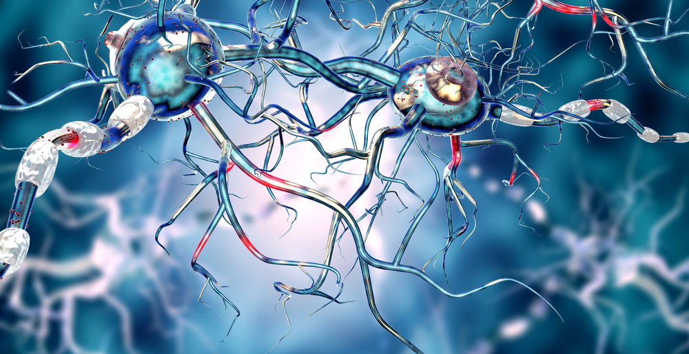
Scientists have identified molecules that enable them to study and manipulate a specific type of cell present in the central nervous system, called glial cells, at the junctions where nerve cells communicate with muscles.
Understanding how these cells operate at such sites, called neuromuscular junctions or synapses, has the potential to allow researchers to understand how certain synapses decay and contribute to disorders like amyotrophic lateral sclerosis (ALS).
Their work, “Specific labeling of synaptic Schwann cells reveals unique cellular and molecular features,” was published in the peer-reviewed journal eLife.
Glia are cells in the central nervous system that do not conduct electrical currents like neurons, but rather help to guide neurons and provide structural support to synapses: the point of contact between two nerve cells that allow them to communicate.
Understanding how synapses develop, and how they are maintained and decay is integral to finding effective therapies for neurodegenerative conditions like ALS.
A significant obstacle to restoring the healthy function of synapses, however, has been an inability to precisely identify the specific types of glial cells that are present in the synapse.
A team led by researchers at Brown University focused on perisynaptic Schwann cells (PSC), a type of specialized, non-myelinating, synaptic glia of the neuromuscular junction (NMJ), that participate in synapse development, function, maintenance, and repair.
Conducting their study in mice, the researchers discovered that these Schwann cells at the neuromuscular junction express, or produce, two proteins: S100-beta and NG2. These proteins unambiguously distinguish PSCs from neighboring cells, much like a “bar code” identifies a household product, Gregorio Valdez the study’s lead investigator, said in a news release.
Other cells express these proteins as well, but only the PSCs found at NMJs expressed both of these proteins together.
Researchers monitored the expression of these two proteins over the mice’s lifespans, evaluating their use in tracking neuromuscular junction development.
They observed that PSCs expressed S100-beta, but not NG2, during embryonic development. NG2 began to appear upon birth, and cells expressing both markers at neuromuscular junctions sharply increased over the first nine days after birth. This coincided with the period of NMJ maturation in mouse skeletal muscles — those that are attached to bones.
By day 21, when neuromuscular junction maturation is nearly complete, the only PSCs observed to express both S100-beta and NG2 were those at the NMJs.
The ability to distinguish one type of cell from another makes it possible to isolate them, and to identify the genes that each cell type preferentially expresses. In PSCs, the Brown team discovered 567 genes enriched in PSCs that were not previously known to be associated with these cells, other glial cells, or synapses.
The “bar code” identified provides scientists with a tool to understand how PSCs contribute to the neuromuscular junction’s repair following injuries, the team suggested, and to its degeneration during both normal aging and the progression of neuromuscular diseases, such as ALS and spinal muscular atrophy (SMA).
“What this means is that we can finally figure out how all three cellular constituents of the synapse — neurons, muscle and glia — talk to each other,” Valdez said. “We now have a unique and important tool for identifying this critical component of the synapse. This is essential for knowing when and where to target to ensure synapses function appropriately.
“This discovery will serve as a springboard to addressing fundamental questions and developing assays to speed the discovery of therapeutics intended to preserve and restore the normal function of neuronal circuits,” Valdez added.


Leave a comment
Fill in the required fields to post. Your email address will not be published.