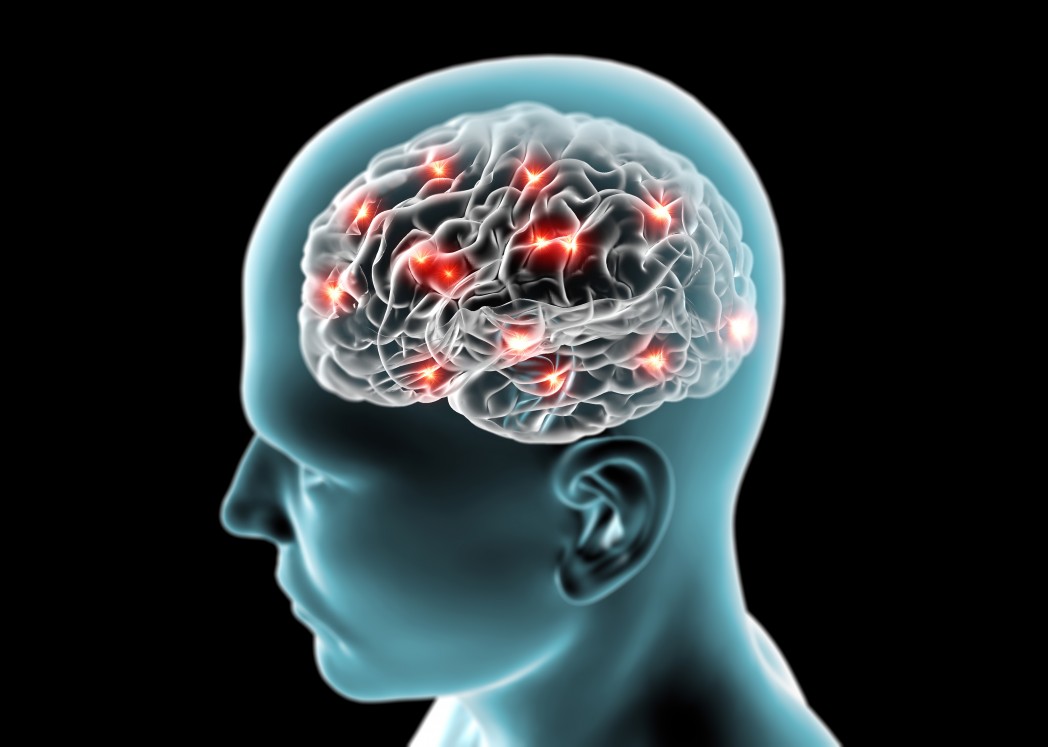
Neuroimaging Techniques Confirm Neuropathological Findings that Contribute to Degeneration in ALS

Advanced neuroimaging techniques and neuropsychological evaluations of patients with amyotrophic lateral sclerosis (ALS) recently confirmed the existence of a unique micro-structural brain degeneration pattern that contributes to memory loss, according to a study conducted by an international research team that included a group from Baylor College of Medicine.
The report, “Memory-related white matter tract integrity in amyotrophic lateral sclerosis: an advanced neuroimaging and neuropsychological study,” was published in Neurobiology of Aging.
The scientists focused specifically on brain areas associated with memory: white matter tracts such as the perforant pathway zone (PPZ), uncinate fasciculus, and fornix.
The researchers enrolled 42 patients with ALS and 25 control participants without the disease who were age- and gender-matched to the patients. Each patient went through specific imaging scans using a 3T MRI, which has a stronger magnetic field and provides clearer images at a faster processing time than standard MRI machines.
Memory tests were also conducted. Comparisons were done to determine whether structural changes in the brain could be correlated with memory-related changes — and if one could cause the other.
The memory testing aspect of the study protocol included the Rey Auditory Verbal Learning Test (RAVLT) to evaluate short-term auditory-verbal memory and retention of information; the Babcock Story Recall Test (BSRT), that asks participants to recall certain aspects of the story after it was read to them; and Rey’s Complex Figure Test (RCFT) which asks participants to draw a complicated line — first by copying it and then by reproducing it from memory.
After analysis of both the imaging and memory testing, the research findings included:
- gender and IQ were contributing variables to the differences found in answers to the RAVLT;
- structural changes found by the imaging testing and gender contributed to the differences observed in answers to the RAVLT immediate recall test;
- structural changes found by the imaging testing contributed to differences observed in answers to the Babcock Story Recall Test-Immediate and Delayed Recall.
“Advanced neuroimaging techniques verified in this study previously reported neuropathological findings regarding PPZ degeneration in ALS. We also detected a unique contribution of microstructural changes in hippocampal and frontotemporal white matter tracts on patients’ memory profile,” the researchers wrote in the study report.
Authors highlighted the need for further conformational studies that would include a larger sample size.


