
Researchers Offer New Insights into Brain Damage in ALS
Written by |
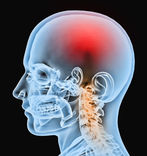
The pathogenesis of amyotrophic lateral sclerosis (ALS) is still not very well understood. A new study on the genetic origin of ALS entitled “Antisense Proline-Arginine RAN Dipeptides Linked to C9ORF72-ALS/FTD Form Toxic Nuclear Aggregates that Initiate In Vitro and In Vivo Neuronal Death” was published in the Journal Neuron by Xinmei Wen part of Dr. Davide Trotti’s group from Frances and Joseph Weinberg Unit for ALS Research, Department of Neuroscience, Thomas Jefferson University, Philadelphia, USA, and colleagues. In this study, the research team found that a frequent gene mutation in ALS produces a protein that seems to be involved in the damaging process of the brain that contributes to ALS.
In amyotrophic lateral sclerosis (ALS), on average 5% of ALS patients have a modified form — expanded GGGGCC (G4C2) nucleotide repeats — of the gene called C9orf72, the most common genetic mutation also associated with frontotemporal dementia (FTD). The normal form of the gene was described in 2011 and since then researchers have been studying its function and the role of the mutated form in the development of ALS.
Dr. Davide Trotti, Ph.D., co-director of the Weinberg Unit, has addressed 3 main hypotheses to explain the function of the C9orf72 mutation in ALS. The first was that the C9orf72 mutation in ALS impaired the normal function of the gene in the cell, but this hypothesis was not right. To test this, the research team inhibited the expression of the gene, using a molecular genetics approach, decreasing the levels of the synthesized protein, but neurons performed normally, suggesting that C9orf72 gene was not crucial to the neuron’s normal function. The second and third hypotheses were that the expanded GGGGCC (G4C2) nucleotide repeats in the gene produced RNA molecules with an anomalous folding structure called a G-quartet, or abnormal folded proteins that were lethal to the cell.
The research team synthesized RNA G-quartets, the abnormal structure of the RNA molecules, and introduced them into normal cells without the C9orf72 mutation. They observed that neurons with longer forms of the quartets died 2 times more than those with less G-quartets, thus suggesting that these abnormal RNA molecules might have a role. However, the most convincing result came when the authors used the proteins originated from the abnormal C9orf72 RNA. They found that one of the five proteins produced from those RNA molecules induced the most lethal effect to the cell. The authors observed that this protein had a chain made from repeats of the amino acids proline (P) and arginine (R), called a poly-PR chain. By following the fate of a living neuron in real-time they could observe that the proteins deposited in the nucleolus and killed the neuron within 72 hours. Next, the authors tested if these processes were also observed in human’s cells. For this, they used iPS cells derived motor neurons from ALS patients to assess the existence of the mutated poly-PR chain C9orf72A protein. In fact, these neurons had the toxic mutated protein. Moreover, when the RNA chains and the PR proteins were both present in the cell, they induced a synergistic effect, suggesting both processes may be involved in causing the damaging effect to motor neurons.
“These studies suggest that if we could prevent the formation of PR aggregates or promote breaking them up, we could help prevent the motor neuron damage that causes the symptoms we see in ALS patients,” said Dr. Trotti, in a press release.


Leave a comment
Fill in the required fields to post. Your email address will not be published.