ALS Patients Exhibit Reduced Cortical Volume, Thickness, and Cerebral Metabolism


 In trying to determine the basis for volumetric changes seen in patients with amyotrophic lateral sclerosis (ALS), Drs. Venkateswaran Rajagopalan and Erik Pioro of the Cleveland Clinic have used voxel based morphometry to measure motor and extramotor grey matter volumes. The two recently delved into a study, titled “Comparing Brain Structural MRI and Metabolic FDG-PET Changes in Patients with ALS-FTD,” and called it, “‘The Chicken or the Egg?’ Question.” Their work was published at the end of 2014 in Journal of Neurology, Neurosurgery & Psychiatry.
Difficulty arises in determining why reduced grey matter volume measurements are seen in patients with ALS due to the means by which the measurement is calculated. VBM analysis counts cortical gyri and sulci as one grey matter region, where gyri are ridges between the sulci valleys in the brain and give the brain its wrinkled appearance. High grey matter volumes can arise from this fact and are sometimes considered erroneous.
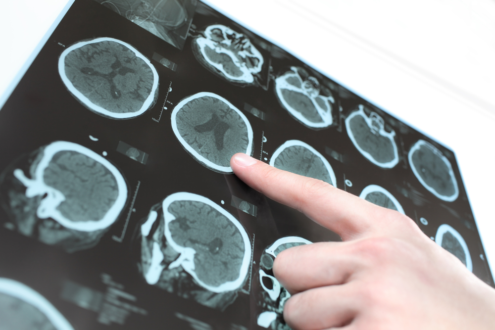
Consequently, Drs. Rajagopalan and Pioro conducted a study to determine if cortical thickness, area, or both contributed to the volumetric changes of grey matter in ALS patients. To do so, the team used T1-weighted MRI in both healthy participants, and ALS patients with frontotemporal dementia (ALS-FTD). The ALS-FTD patients also underwent positron emission tomography (PET) to determine metabolic changes in the measured structures.
MRI images revealed significant reductions in grey matter volume and cortical thickness in ALS-FTD patients when compared to healthy controls. Conversely, there was no significant reduction in cortical surface area in any of the examined brains. Therefore, the researchers determined, “Significant reductions primarily in cortical thickness were the likely reason for decreased grey matter volume in ALS-FTD.”
Within any region that saw a significant structural change, there was a concomitant reduction in cerebral glucose metabolism, as determined by PET. “Metabolic changes corresponded well with structural changes in motor and extramotor areas, and sometimes occurred even in the absence of grey matter volume reduction,” wrote the authors. Moving forward, the authors believe their study suggests that neurodegeneration can be considered a “neuronopathy” in the case of ALS-FTD.