Specially Engineered Neural Cells Delay Disease and Extend Life in ALS Animal Model

Transplanting engineered neural cells into the brain of an amyotrophic lateral sclerosis (ALS) animal model delayed disease progression and extended the animals’ survival, a study shows.
The study, “Transplantation of Neural Progenitor Cells Expressing Glial Cell Line‐Derived Neurotrophic Factor into the Motor Cortex as a Strategy to Treat Amyotrophic Lateral Sclerosis,” was published in the journal Stem Cells.
Currently available therapies are only capable of managing ALS symptoms, such as muscle spasms, but there is no treatment that can reverse the damage or cure the disease. As a result, the prognosis for ALS patients is poor.
“If we are able in the future to reproduce our research results in humans, we could improve both the quality and length of life for patients diagnosed with this devastating disease,” Gretchen Thomsen, PhD, a research scientist at the Cedars-Sinai Board of Governors Regenerative Medicine Institute and the study’s first author, said in a press release.
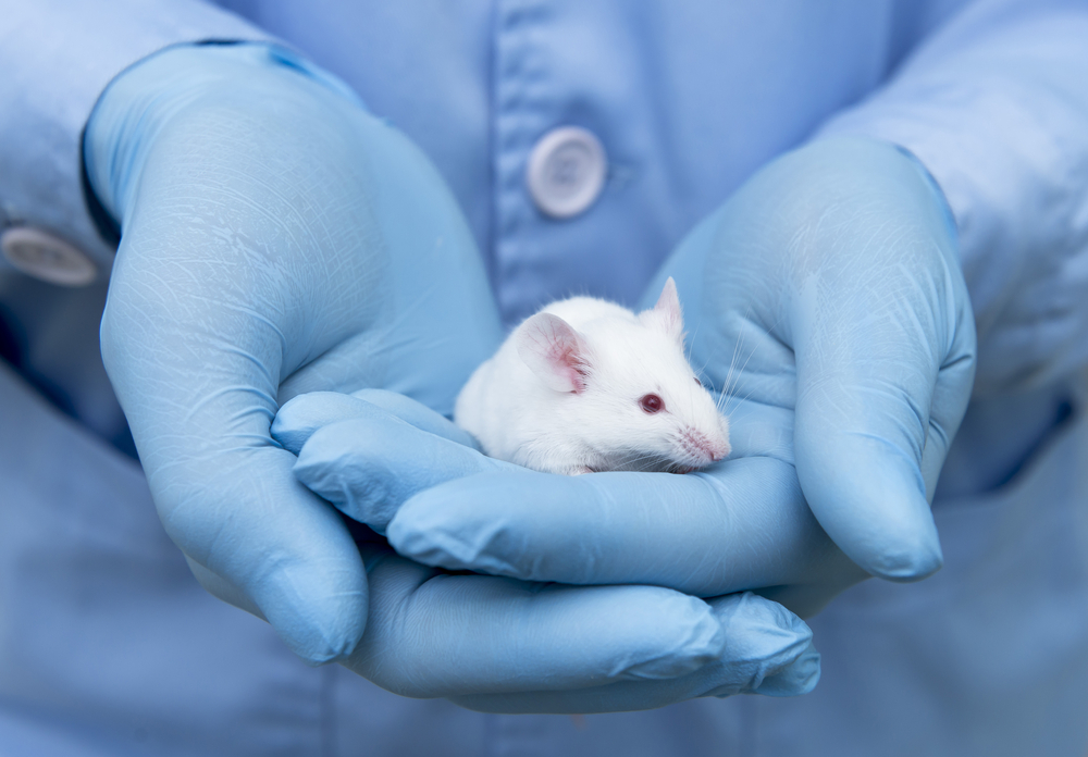
Researchers at Cedars-Sinai Medical Center investigated the outcomes of transplanting neural progenitor cells engineered to secrete a special protein called GDNF into the brain of a rat model of ALS.
GDNF, short for glial cell line‐derived neurotrophic factor, is a powerful growth factor that was shown to protect motor neurons and help preserve the neuromuscular junctions — the contact point between a motor nerve cell and a muscle fiber – improving the outcomes of ALS animal models.
In this study, researchers saw that once inside the brain’s cortex, the GDNF-secreting cells matured into astrocytes — specialized cells that help support motor neurons — and released GDNF.
The transplanted cells delayed the disease and extended the survival of the animals: Survival was 8 percent higher (median survival was 184 days for the transplanted versus 170 days for the non-transplanted animals), and animals were free of paralysis 10 percent longer than non-transplanted animals (175 days for the transplanted versus 159 days for the non-transplanted animals).
Survival of corticospinal motor neurons — those controlling the muscle movements — was also enhanced in the animals.
Investigators say more preclinical work is now being conducted to determine the safest and most effective treatment levels. Meanwhile, a Phase 1 clinical trial (NCT02943850) is already underway testing the transplanted cells in 18 ALS patients.
The trial, launched in 2016 at Cedars-Sinai, is evaluating the safety of two doses of the GDNF-producing neural progenitor cells when delivered into the spinal cords of patients that have ALS-related weakness in their legs. The study is still recruiting patients.
“Cedars-Sinai is committed to pursuing groundbreaking research that aims to one day eradicate ALS and the profound human suffering of this disease,” said Shlomo Melmed, executive vice president of academic affairs and dean of the medical faculty at Cedars-Sinai. “Today’s study significantly advances that drive.”
Researchers at Cedars-Sinai have long studied ALS: A recent study published in the journal Stem Cell Reports, showed that blood vessels in the brain can activate genes promoting spinal motor neurons to grow; and a 2016 study in the journal Science found that immune cells in the brain may play a direct role in ALS development.


