ALS MRI Marker May Facilitate Diagnosis and Monitoring, Study Finds


A magnetic resonance imaging (MRI) marker may facilitate amyotrophic lateral sclerosis (ALS) patients’ diagnosis and disease monitoring, a study finds.
The study, “Is the Hypointensity in Motor Cortex the Hallmark of Amyotrophic Lateral Sclerosis?,” was published in the Canadian Journal of Neurological Sciences.
ALS is a progressive neurological disorder in which motor neurons — the nerve cells responsible for controlling voluntary muscles — gradually degenerate and die, causing muscles to shrink (atrophy) and become weaker. Besides lack of muscle strength, more than half of ALS patients also experience cognitive impairments over the course of the disease.
The clinical variability of ALS symptoms and their similarity with other types of neurodegenerative disorders can make the clinical diagnosis of ALS difficult. Based on MRI data, some studies have proposed that neural degeneration starts affecting upper motor neurons (UMNs) (“dying-forward” hypothesis), while others argued that lower motor neurons (LMNs) (“dying-back” hypothesis) are the first to be affected by the disease.
“There are insufficient strategies for exploring the pyramidal tract [UMNs that connect the brain to the brainstem or spinal cord] to inform early ALS pathology [disease development], underscoring a need for UMN biomarkers,” the researchers said.
After observing that several ALS patients had lower MRI signal in one of the ridges of the brain’s frontal lobe in the shape of a ribbon (the “black ribbon,” or BR), a group of Mexican researchers set out to evaluate the usefulness of this MRI marker as a diagnosis tool for ALS.
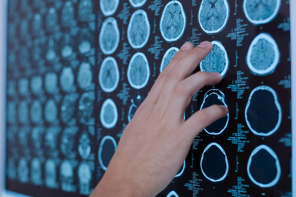
The prospective study involved 36 ALS patients (20 men and 16 women) with a mean age of 54.5 years, and 15 healthy controls (eight men and seven women) with a mean age of 54.4 years. All study participants performed 3T brain MRI scans at the beginning of the study and 18 months later under the same conditions.
As expected, most ALS patients (91.66%) had the BR in the brain’s frontal lobe, covering an average area of 79.98 mm3. MRI signal intensity under the optimal cutoff value of 83 arbitrary units (AU) successfully distinguished ALS patients from healthy controls with 97% accuracy, 92% sensitivity, and 93% specificity.
In addition, correlation analyses showed that loss of muscle strength and respiratory forced vital capacity were directly correlated with MRI signal intensity in BR.
All seven ALS patients who enrolled in a second study at 18 months follow-up showed significant alterations in signal intensity and area in the BR region compared to their first examination, suggesting that changes in BR reflect ALS disease progression.
“In summary, to our knowledge, this study is the first to show such a high sensitivity in an MRI marker of ALS. This sign should be developed in larger groups of patients with other motor neuron diseases at earlier stages and also in longitudinal studies, such as clinical trials, in order to test its reliability,” the scientists concluded.