Tiny Microscope Lets Scientists Peer into Spinal Cords of Mice and See Astrocytes in Action

Researchers using a miniature microscope saw that glial cells, called astrocytes, contribute to sensory nerve transmission in the spinal cord of awake and moving animals. Offering unparalleled insights into the workings of the spinal cord, the tool may lead to new treatments for a range of neurodegenerative diseases, including amyotrophic lateral sclerosis (ALS).
A decade ago, the general consensus in the scientific community was that glial cells like astrocytes were merely passive bystanders to the activities of nerve cells, supporting them with nutrients and structural support. But other researchers around that time began to show that astrocytes, as well as other glia, actively regulate nerve transmissions by releasing their own factors, a process that became known as gliotransmission.
While much research has been done since on the signaling abilities of astrocytes, none were able to observe them in action in conscious animals. Researchers at Salk Institute for Biological Studies, California, have now done just that — and describe their findings in the study, “Imaging large-scale cellular activity in spinal cord of freely behaving mice,” appearing in the journal Nature Communications.
“For a long time, researchers have dreamed of being able to record cellular activity patterns in the spinal cord of an awake animal. On top of that, we can now do this in a freely behaving animal, which is very exciting,” the study’s first author, Kohei Sekiguchi, a PhD student at the University of California, San Diego, said in a news release.
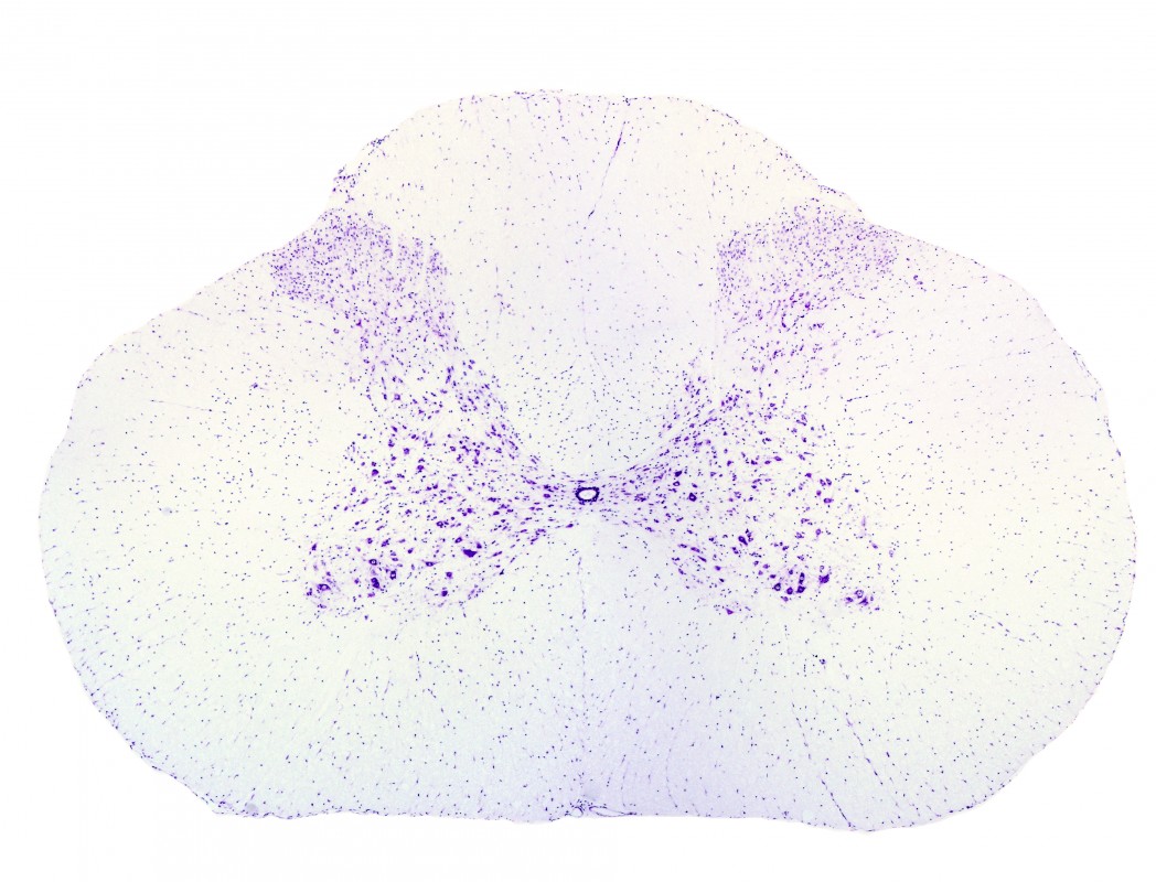
The spinal cord is a hub for nerve connections between the central nervous system and more peripheral body parts. Despite its importance in sensory processing, researchers still don’t know how nerve signals carrying sensory information are encoded in the cells of the spinal cord.
The miniature microscope — an invention the team worked to refine since 2008, and which is about the size of a penny — has previously been used by the team to explore the brain. Investigations of the spinal cord have, however, been hindered by technical difficulties. The spinal cord is surrounded by independently moving vertebra and is close to pulsating organs, making a clear view difficult to achieve.
With an improved technique, Axel Nimmerjahn — the study’s senior author and an assistant professor in Salk’s Waitt Advanced Biophotonics Center — and his team could observe freely moving mice. The researchers noted that different types of sensory stimuli, such as light touch or pressure, did not activate the same subsets of neurons. They also observed how astrocytes reacted to the stimuli in a coordinated manner.
“Not only can we now study normal sensory processing, but we can also look at disease contexts like spinal cord injury and how treatments actually affect the cells,” Dr. Nimmerjahn said.
The team will next attempt to study touch and pain sensory signaling simultaneously in the brain and spinal cord at even higher resolutions.


