Fish Model of ALS Links TDP-43 Protein Clumping to Motor Neuron Ills

A zebrafish model that reproduces key amyotrophic lateral sclerosis (ALS) symptoms and characteristics, including protein clumping, when exposed to blue light could aid in understanding disease mechanisms and in developing new treatments.
The model was described in the study, “Optogenetic modulation of TDP-43 oligomerization accelerates ALS-related pathologies in the spinal motor neurons,” published in Nature Communications.
Nearly all cases of ALS are characterized by the formation of aggregates (clumps) containing the protein TDP-43. These aggregates are believed to be drivers of motor neuron death in the disease. But to study this directly requires a system where TDP-43 clumps only under certain conditions that can be experimentally manipulated.
That is, one would need a model where TDP-43 aggregation could be ‘turned on’ in a way that’s easy to control, so as to see whether such clumping directly causes motor dysfunction.
Researchers used optogenetics to create such a model. This technology involves engineering proteins to be sensitive to certain wavelengths of light.
In this case, they created zebrafish with a light-sensitive version of TDP-43, which they dubbed opTDP-43 (optogenetic TDP-43 variant).
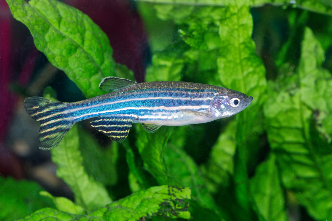
Zebrafish are commonly used to study neuronal conditions because their motor neurons work in ways quite similar to humans. The larvae of these small tropical fish are also transparent, allowing light to be applied to cells in the nervous system with minimal invasiveness.
In the dark, opTDP-43 behaved like normal TDP-43 protein. That is, it was present in the nucleus (the cellular compartment that houses DNA), which is where this protein is normally found. But when the fish were exposed to blue light, the protein moved outside of the nucleus to the cytoplasm, as happens in ALS patients.
Continued exposure to blue light led to the formation of opTDP-43 aggregates after about three days. Interestingly, prolonged exposure (more than four days) led normal TDP-43 to also move to the cytoplasm and form aggregates with the opTDP-43.
“These observations raise a possibility that a long-term illumination turns opTDP-43h [human opTDP-43] into aggregates that incorporate non-optogenetic TDP-43,” the researchers wrote.
When these aggregates were formed, the fish’s neurons were smaller, and muscle innervation (the amount of connections between nerve and muscle cells) lesser, recapitulating what happens in human ALS.
“This research, for the first time, shows that TDP-43 aggregation is a cause of motor neuron dysfunction in animals,” Kazuhide Asakawa, PhD, a professor at Tokyo Medical University and study co-author, said in a press release.
While TDP-43 aggregation happens in nearly all ALS cases, certain mutations in the gene that codes for TDP-43 are associated with familial ALS, presumably because they can exacerbate or predispose a person to this aggregation.
The researchers created a version of opTDP-43 with such a mutation (A315T). They found that, under exposure to blue light, the protein also formed aggregates, and this led to more pronounced motor problems than were seen in the non-mutated opTDP-43 protein.
Further experimentation suggested that this was because the mutant TDP-43 was more stable in the cytoplasm, helping to facilitate the stable formation of aggregates.
Overall, this study describes a new animal model of ALS that could be important for research into the disease.
“We can now produce an ALS-like state in a temporary and spatially tuned manner by controlling light intensity and the position of illumination,” Asakawa said. “Our ultimate goal over the next few years is to identify chemicals that prevent optogenetic TDP-43 from forming oligomers and aggregates, and we hope such chemicals will be used for ALS treatment.”


