
New Cellular Model Used to Study FUS Gene Mutations
Written by |
Mutations in the FUS gene, a common cause of amyotrophic lateral sclerosis (ALS), impair the regeneration and growth of motor neuron extensions toward muscle cells, according to a study using a new miniaturized human model of neuron-muscle interactions.
These deficits, along with the reduced number of nerve-motor connections, were lessened by blocking the HDAC6 enzyme, supporting previous studies pointing out HDAC6 suppression as a potential therapeutic strategy for ALS.
Selective, brain-penetrant HDAC6 suppressors are being developed by Eikonizo Therapeutics, in collaboration with the McQuade Center for Strategic Research and Development, for neurodegenerative diseases such as ALS.
The findings also support the use of this human-derived model for studying the effects of ALS-associated mutations in the motor neuron-muscle connections and the effectiveness of potential therapies for ALS, the researchers noted.
The study, “Human motor units in microfluidic devices are impaired by FUS mutations and improved by HDAC6 inhibition,” was published in the journal Stem Cell Reports.
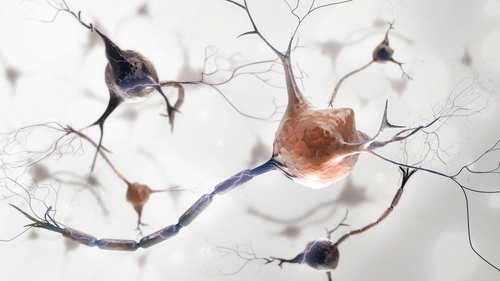
The neuromuscular junction (NMJ) is the site of communication between motor neurons — the specialized nerve cells that control voluntary muscle and that are gradually lost in ALS — and muscle cells. This communication is driven by long motor neuron fibers, called axons, that send chemical signals to the muscle.
There is “ample evidence that the disconnection of the motor axon from the muscle is the first step in the disease process of ALS, making it an important mechanism to study,” the researchers wrote.
Studying NMJs and their relation to ALS may help to better understand the neurodegenerative disease and identify potential therapeutic targets and strategies. Yet, a standardized, accessible, human cell-derived model of these NMJs that allow the maintenance of cell type-specific microenvironments, is lacking.
Now, a team of researchers at the University of Leuven, in Belgium, generated such a human-derived model using microfluidic devices, which have tiny chambers to grow cells in which fluid is passed through to mimic fluid flow in a whole organ.
In this case, the device contained two separate mini-chambers on opposite sides, one growing motor neurons and the other muscle cells, connected through tiny channels.
The team first found that, using cells derived from healthy individuals, the new system enabled the motor neurons to grow their axons through the tiny channels and reach muscle cells in the other chamber, forming functional NMJs — thereby validating the model.
The researchers then evaluated NMJ dynamics in the same model, but using motor neurons derived from two ALS patients carrying distinct FUS mutations. These were compared with corresponding patient-derived cells genetically corrected for those mutations (used as controls).
Mutations in the FUS gene are present in about 5% of familial ALS cases and about 1% of sporadic ALS cases, and may lead to some of the most aggressive forms of ALS, including a type that begins during adolescence and young adulthood.
FUS mutations can result in the production of an abnormal FUS protein that is prone to form toxic aggregates, which can build up over time and lead to nerve cell damage.
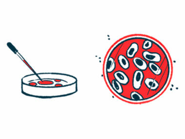
Patient-derived motor neurons were generated via induced pluripotent stem cells (iPSCs) engineered from patients’ own skin cells, which were reprogrammed back to a stem cell-like state, where they can give rise to almost every type of human cell.
As such, iPSCs can be genetically modified to carry known disease-causing mutations or derived directly from patients to be used as cellular models that mimic the disease’s genetic and clinical diversity.
Results showed that FUS-mutant motor neurons grew fewer axons between the chambers and formed significantly fewer (nearly half) NMJs with muscle cells in the neighboring chamber, when compared with respective control motor neurons.
In addition, patient-derived motor neurons with FUS mutations regenerated damaged axons less efficiently.
These findings highlighted that “the number of NMJs and the [motor neuron axon] outgrowth/regrowth are affected by mutations in FUS,” the researchers wrote, adding that “these impairments can be successfully rescued by correction of the mutations, emphasizing that the effects are a direct cause of [FUS mutations].”
Notably, the team’s previous work showed that blocking the activity of histone deacetylase 6 (HDAC6), an enzyme involved in molecule transport within cells, rescued axonal transport defects in motor neurons derived from people with FUS-associated ALS.
As the observed impairments in axon growth and repair may be related to axonal transport problems, the researchers investigated whether treating the cellular model with Tubastatin A, a widely used HDAC6 suppressor in preclinical studies, could also restore these deficits.
The team found that Tubastatin A improved FUS-mutant axonal growth and post-injury regrowth to levels similar to controls and increased the number of NMJs formed.
Notably, Tubastatin A only restored axon regrowth when administered after injury, and not before, suggesting that “HDAC6 inhibition could be useful as a treatment after injury rather than a [preventive] therapy,” the researchers wrote.
Overall, the new cellular model is a versatile, valuable, and relatively easy-to-use tool “to assess human NMJ physiology in health and disease” and “can be used to test new therapeutic strategies for [motor neuron] diseases,” the team added.
Future studies in animal models, and ultimately in ALS patients, are needed to further clarify the therapeutic potential of blocking HDAC6 in ALS. Notably, Tubastatin A has limited brain penetrance, making it a poor candidate for ALS, but brain-penetrant HDAC6 suppressors are currently under development by several academic groups and pharmaceutical companies.


Leave a comment
Fill in the required fields to post. Your email address will not be published.