
Human Cell Model Captures Brain and Muscle Interaction for 1st Time
Written by |
A system using human cells to model how the brain controls muscle movement has been developed for the first time, and scientists expect it to aid in understanding neurological disorders that affect movement, including amyotrophic lateral sclerosis (ALS).
The study describing their model system, “Generation of Functional Human 3D Cortico-Motor Assembloids,” was published in Cell.
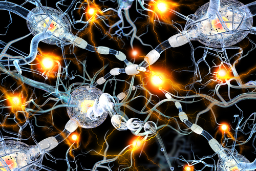
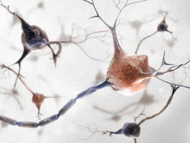
From a neurological perspective, voluntary movement can be broken down into three general steps: an electrical signal originates in the brain, and then is carried along the spinal cord. When the signal arrives at muscles, it prompts them to contract, resulting in movement.
ALS is characterized by the death of the nerve cells involved in motor movement, through mechanisms that are still not completely understood. Studying animal models like mice can provide some insights into the disease; however, the nervous systems of rodents and humans have fundamental differences that hamper research efforts.
“ALS has been cured in rodents dozens of times. None of the cures have effectively translated to people,” Sergiu Pasca, MD, a professor at Stanford School of Medicine, and a neuroscientist and study co-author, said in a press release.
To address the limitations of animal models, Pasca and colleagues at Stanford and BD Biosciences developed a model of the nervous system that uses human cells.
Specifically, the researchers used human induced pluripotent stem cells (iPSCs), a type of stem cell that is generated from mature cells, typically skin or blood cells, using specialized techniques. Being “pluripotent” means that iPSCs can become many other types of cells, depending on the particular chemical cues they are given.
By treating iPSCs with specific combinations of signaling molecules, the research team differentiated the iPSCs into populations of cells called spheroids. These three-dimensional cellular structures more closely mimic the layout of cells in a human body than do the more traditional use of cells in dishes.
In prior work, the Stanford researchers created spheroids that closely mimic the architecture and physiology of the cerebral cortex, a part of the brain that includes a region which controls voluntary movement. In this new work, the researchers adjusted their protocol to create two additional types of spheroids: one that mimics the spinal cord, and another that mimics muscles.
When these three types of spheroids were placed next to each other in a dish, they combined into structures called assembloids. Effectively, they formed a cellular model of the brain, connecting to the spinal cord, connecting to muscle. A battery of experiments demonstrated that these circuits had accurately assembled themselves.
“We made the parts, but they knew how to put themselves together,” Pasca said.
The assembloids were shown to be functional, as stimulating the “brain” part of the assembloids caused a twitch in the muscle component.
“Skeletal muscle doesn’t usually contract on its own,” Pasca said. “Seeing that first twitch in a lab dish immediately after cortical [brain] stimulation is something that’s not soon forgotten.”
The assembloids can also be effectively kept in culture for up to 10 weeks. “Remarkably, the longer they [the assembloids] remain intact, the better the contraction is,” Pasca said.
“This feature may allow for further refinement and maturation of connections,” the researchers wrote. They speculated that such long-term culture may allow for myelination — the formation of the myelin sheath, a fatty substance around neurons that is necessary for efficient electrical signaling, and is damaged in many neurological disorders.
“There are a number of potential applications for this cellular platform that could be used to gain insights into the evolution, development, and disorders of the cortico-spinal-muscle circuit,” the researchers wrote.
Their model could be used to better understand how the human nervous system works, both in people with good health and those with diseases like ALS, they noted. Incorporating other types of cells into the model, like immune cells, could also be useful for understanding conditions like multiple sclerosis, where the immune system attacks the nervous system.
“Ultimately, assembloids of various parts of the [nervous system] could bring insights into understanding assembly of different types of human circuits and into identifying therapeutic strategies,” the researchers concluded.

Leave a comment
Fill in the required fields to post. Your email address will not be published.