New Relevant Mouse Model for the Study of ALS and Frontotemporal Dementia
Written by |

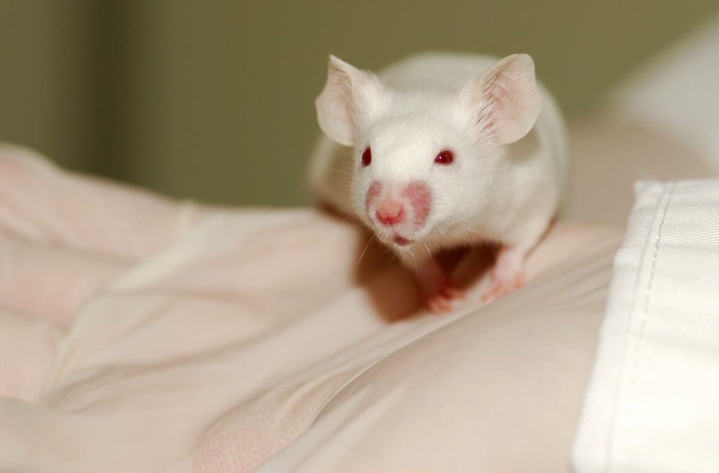
A new study led by researchers at the Mayo Clinic and recently published in the journal Science reported a new, important mouse model that allows the study of amyotrophic lateral sclerosis (ALS) and frontotemporal dementia, two relevant neurodegenerative diseases. The study is entitled “C9ORF72 repeat expansions in mice cause TDP-43 pathology, neuronal loss, and behavioral deficits.”
The ALS Association estimates that more than 300,000 Americans suffer from ALS, a progressive neurodegenerative disease characterized by the gradual degeneration and atrophy of motor neurons in the brain and spinal cord that are responsible for controlling essential voluntary muscles, such as the ones related to movement, speaking, eating and even breathing. ALS patients may become totally paralyzed and the majority die due to respiratory failure within two to five years post diagnosis.
Frontotemporal dementia refers to a group of disorders that mainly affect the frontal and temporal lobes of the brain, which are associated with personality, behavior and language. Depending on the brain area affected, the symptoms may vary. Some patients experience dramatic changes in their personality, while others lose the ability to use language. This condition is often misdiagnosed as Alzheimer’s disease or a psychiatric disorder. The Alzheimer’s Association estimates that frontotemporal dementia accounts for up to 10 – 15% of all diagnosed dementia cases.
It is known that the major genetic cause of both ALS and frontotemporal dementia is a repeat expansion in the gene C9ORF72. Advances in the study and treatment of these disorders have been hampered by the lack of a suitable animal model that can mimic the features of the diseases. Researchers have now developed a mouse model able to mimic the clinical, behavioral and neuropathological characteristics linked to both ALS and frontotemporal dementia.
“Our mouse model exhibits the pathologies and symptoms of ALS and FTD seen in patients with the C9ORF72 mutation,” noted the study’s senior author Dr. Leonard Petrucelli in a news release.
Using this mouse model, researchers found that C9ORF72 mutation induces the production of toxic RNA species in the brain, forming aggregates of C9RAN protein along with TDP-43 protein, a protein previously reported to be deregulated in ALS and frontotemporal dementia.
“Finding TDP-43 in these mice was entirely unexpected—and we could not have discovered a link between the repeat expansion in C9ORF72 and development of TDP-43 pathology without the mouse model,” noted Dr. Petrucelli. “We don’t yet know how foci and c9RAN proteins are linked to TDP-43 abnormalities or what the pathway is, but with our new animal model, we now have a way to find out.”
The team suggests that the changes found in the mouse brain could account for the behavioral and motor deficits observed in human patients. At six months of age, mice expressing the expanded C9ORF72 repeat in their central nervous system were found to exhibit anxiety, hyperactivity, motor deficits and antisocial behavior.
The team’s next goal is to identify chemical compounds that are able to bind to the repeat-expansion RNA prior to aggregate formation. The idea is that targeting of the RNA will potentially inhibit the formation of RNA aggregates and c9RAN proteins in brain cells, and therefore prevent nerve cell function impairment. The authors believe that this mouse model can offer a new tool to test potential chemical compounds and therapeutic agents against nerve cell death.


