Mice with ALS-like Disease Function Better and Live Longer When Protein Receptor’s Activity Is Reduced

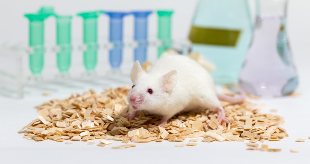
Researchers have slowed the progression of an ALS-like disease in mice, and prolonged their lives, by reducing the activity of a protein receptor that helps transmit signals between nerve cells.
The study, which involved the metabotropic glutamate receptor 5, or mGluR5, was published in the journal Neuropharmacology. The article was titled “In-vivo effects of knocking-down metabotropic glutamate receptor 5 in the SOD1G93A mouse model of amyotrophic lateral sclerosis.”
ALS is caused by loss of motor neurons, or nerve cells that control muscles. Scientists are uncertain what causes them to die. But one possibility is neurotoxicity, or over-stimulation of motor neurons by substances such as neurotransmitter glutamates, which carry signals between nerve cells.
MGluR5 is a protein found on the surface of neurons. It occurs at the neurons’ synapses, or junctions between the cells where the transmission of signals takes place.
The function of the protein, which is found on neurons in specific parts of the brain, is to take up glutamate and increase the synapse’s excitability, or responsiveness.
Researchers halved the activity of mGluR5 receptors in mice with an ALS-like disease by breeding out of the animals one of two genes that code for the mGluR5 protein. This led to a more normal glutamate release in the mice’s spinal cords.
Signs of amyotrophic lateral sclerosis appeared later in the mice, and the animals survived longer than mice whose mGluR5 activity was not reduced.
In addition, more motor neurons remained healthy and survived in the mice with the diminished mGluR5 activity. And two kinds of brain cells, astrocytes and microglia, which the body activates when the brain experiences injury, showed less activity in the test mice than in the untreated mice.
Another thing that researchers discovered in the mice with half the mGluR5 activity was less calcium in the cytoplasm of the animals’ neuron cells. Calcium plays a role in transmitting signals between neurons.
The movement functioning of the male mice improved, although in the females it did not, researchers said.
The same group of researchers obtained similar, but less striking, results when they halved the expression of a related glutamate receptor, mGluR1, in mice with ALS. That finding led to a different article, “Knocking down metabotropic glutamate receptor 1 improves survival and disease progression in the SOD1G93A mouse model of amyotrophic lateral sclerosis.”
“The present results obtained by reducing mGluR5 expression in the mouse model of ALS, along with previous evidence targeting mGluR1, emphasize the role of Group I mGluRs in the development of ALS and allows proposing the hyper activation of these Glu receptors as a novel mechanism contributing to the disease progression,” the researchers wrote.
“The reduced expression of mGluR5 determined delayed clinical onset of the disease, amelioration of symptoms, MN (motor neuron) preservation, and improved biochemical and functional readouts. Our results suggest that pharmacological treatments aimed at blocking Group I mGluRs can reasonably turn out to be effective in counteracting ALS.”


