Transcranial stimulation seen to boost muscle strength, life quality
Additional treatment courses in small trial also extend patient survival by 71%

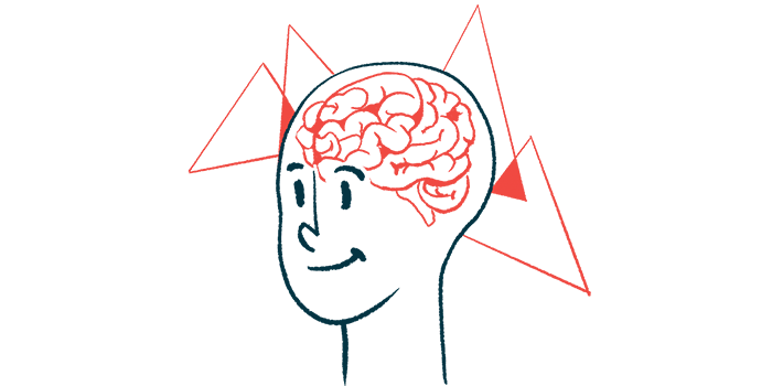
Transcranial stimulation — a noninvasive technique that delivers low electrical currents to the brain to fine-tune nerve cell activity — improved muscle strength and quality of life in people with amyotrophic lateral sclerosis (ALS) in a small clinical trial.
Researchers also found that a two-week course of transcranial direct current stimulation (tDCS) restored electrical activity through certain neuronal circuits, and that each additional treatment course significantly lowered the risk of death by 71%.
“Our study provides compelling evidence for the potential benefits of cortico-spinal tDCS as a therapeutic and rehabilitative approach for patients with ALS,” the scientists wrote. Still, “larger and more comprehensive clinical trials are necessary to confirm these results and further investigate the mechanisms underlying these effects.”
The study, “Cortico-spinal tDCS in amyotrophic lateral sclerosis: A randomized, double-blind, sham-controlled trial followed by an open-label phase,” was published in Brain Stimulation.
Transcranial stimulation allows neurons to fine-tune responses to stimuli
ALS is a progressive disease that damages motor neurons, the nerve cells that control voluntary muscles in the body. Without motor neurons, the brain is no longer able to control movement, causing muscle weakness and a range of other disease symptoms.
Life expectancy with ALS varies depending on which symptoms appear first and how rapidly the disease progresses. While medications can slow disease progression and extend survival, not every patient responds well to current disease treatment options.
Transcranial direct current stimulation is a noninvasive approach that uses constant, low-intensity electrical currents, passed through small electrodes applied to the scalp, to stimulate certain regions of the brain.
It aims to fine-tune neuronal responses to stimuli, allowing the brain to rewire itself in a way that differs from how it previously functioned. This has been shown to slow progression in neurodegenerative diseases such as Parkinson’s, and it is thought to hold promise in ALS.
Researchers in Italy conducted a two-part clinical trial (NCT04293484) to assess tDCS in 31 adults, all with a disease diagnosis of early to moderate stage ALS, at a single site in Brescia. Patients in its first part were randomly assigned to either sham (placebo) stimulation or tDCS for five days each week over two weeks.
Using a so-called cortico-spinal approach, scientists stimulated both the motor cortex — a brain region involved in controlling voluntary muscle movement — and the spinal cord at the same time.
“In ALS, cortical hyperexcitability has emerged as a key factor contributing to the degeneration of motor neurons,” the researchers wrote.
In the study’s second part, both patient groups received tDCS about six months after the first course. Follow-up evaluations were done up to 48 weeks from baseline (the study’s start).
Significant gains in muscle strength seen, starting with two weeks of treatment
The trial’s main goal was to determine whether tDCS could increase muscle strength, as assessed with the Medical Research Council (MRC) scale, which ranges from 0 (no movement) to 5 (normal contraction). This score is the sum of scores for each muscle group, like shoulder abductors and knee flexors, with total scores ranging from 100 (no impairment) to 0 (most severe impairment).
Compared with trial’s sham stimulation, tDCS use resulted in significant improvements in muscle strength at week 24 (about six months), with most improvements seen during the two weeks of treatment. While scores rose by an average of 0.9 points on the MRC scale for people on tDCS, these scores dropped by 6.6 points on average in the sham group.
Treatment also resulted in a better quality of life for patients and a lower burden for caregivers over the 24 weeks.
Nineteen patients had data on intracortical connectivity, or how neurons communicate with each other through circuits within the brain’s cortex. tDCS significantly improved connectivity processes known to be disturbed in people with ALS.
The treatment also appeared to lessen neuronal damage in patients, as assessed by changes in the levels of neurofilament light chain (NfL), a nerve damage biomarker.
While NfL levels peaked about eight weeks after the start of tDCS, they markedly dropped after that, reaching the lowest levels at the 48-week assessment. For initial sham group patients who began tDCS treatment at study week 24, reductions in NfL levels started to become evident between 26 and 32 weeks.
By the study’s end, 15 patients had died. Bulbar-onset disease, which initially affects the muscles around the face and throat, causing slurred speech and difficulty swallowing, linked with a nearly five times higher risk of death.
In turn, for each tDCS course, there was a 71% lower risk of death, “suggesting that the number of completed tDCS treatments significantly reduced the risk of death and increased survival,” the researchers wrote.
“Our study presents several encouraging findings regarding the therapeutic potential of cortico-spinal tDCS for patients with ALS, comprehensively encompassing highly relevant aspects of ALS: global muscle strength, patient-rated quality of life, caregiver burden and survival,” the researchers concluded.
Despite its small number of patients, “the promising results from our study highlight the potential of cortico-spinal tDCS as a therapeutic intervention for ALS patients, and they open up several avenues for future research,” they added.


