New System Mimics Human Testing of Therapies for Neuromuscular Junction Diseases Like ALS


New technology that tests therapies designed for disorders of the neuromuscular junction, such as amyotrophic lateral sclerosis (ALS), closely mimics the human condition and offers a new method of drug testing, according to a report.
The paper, “Stem cell derived phenotypic human neuromuscular junction model for dose response evaluation of therapeutics,” was written by Hesperos Chief Scientific Officer James Hickman, PhD, funded by the National Institutes of Health, and published in the journal Biomaterials.
Hickman is a professor of chemistry, biomolecular science, and electrical engineering at the Hybrid Systems Laboratory at the University of Central Florida.
The new technology is licensed to Hesperos, and is part of the company’s human-on-a-chip program, which re-creates specific human systems in the laboratory for the purpose of toxicology testing and evaluating the effectiveness of new therapies. The new system is available as a fee-for-service assay.
The most common method of testing any new therapy before human clinical trials is animal testing. But animal tests can be inaccurate because only one out of 50 experimental treatments that are safe in animals are found to be safe in humans.
As a result, there is a need for more systems that can more properly mimic human biological processes.
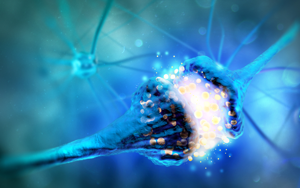
The neuromuscular junction (NMJ) is where motor neurons — cells of the nervous system — meet muscle cells. The two cell types communicate with each other by releasing molecules into the NMJ, which then bind to the other cell type.
Diseases characterized by motor impairment, such as ALS and spinal muscular atrophy (SMA), have an impairment of the NMJ.
There are currently no NMJ systems made of human cells that can be used for drug testing.
And there are no model systems — animal or human — that allow for isolated treatment of motor neurons or muscle cells for researchers to determine the effectiveness of a therapy in these highly specialized cells.
With this in mind, researchers developed a system in which human muscle cells and motor neurons derived from stem cells are cultured in a two-chamber BioMEMS construct that allows separate stimulation of each component.
BioMEMS — short for biomedical microelectromechanical systems — are medical microdevices that are used for biomedical research.
Over time, muscle fibers called myotubes will form from muscle cells, and the motor neurons will form neuromuscular junctions with the myotubes.
The NMJ model encapsulates a setting in which drug testing re-creates real evaluation conditions. It allows for the administration of therapies in either a single or several doses over an extended time period.
Researchers used this system to test the dose response of three different therapies: curare toxin, alpha bungarotoxin, and botulinum toxin (Botox).
The dose response results of the treatments from the NMJ model matched those of live human data, suggesting that it can allow for realistic testing of pharmacological therapies.
“The model’s sensitivity in quantifying the degree of loss-of-function caused by neuromuscular blocking agents with varying modes of action provides a highly accurate and sensitive screening tool for new drugs,” Hickman said in a press release.
Because the system can be created using stem cells from patients, it can be used to generate patient-specific NMJ models.
“It can also allow us to observe the behavior of neuromuscular systems as a disease progresses, and inform treatment decisions based on what patients are experiencing, as they experience it,” Hickman said.