Boosting Protein with Role in Getting Nutrients to Cells Prevented Muscle Wasting in ALS Mice, Study Finds


Restoring or artificially raising levels of a protein found in the spinal cord, mitofusin 2, worked to prevent nerve degeneration, muscle atrophy, and paralysis in a mouse model of amyotrophic lateral sclerosis, researchers report. As this protein is often depleted in ALS patients — and drops with aging — they believe that supplementing it offers a way of preserving skeletal muscle health.
These findings were reported in the study, “Mitofusin 2 Regulates Axonal Transport of Calpastatin to Prevent Neuromuscular Synaptic Elimination in Skeletal Muscles,” published in Cell Metabolism.
ALS is a progressive neurological disorder that destroys motor neurons — the nerve cells responsible for controlling voluntary muscles — to cause extreme physical disability. Without these cells, the brain is no longer capable to control muscle movement, and muscles shrink (atrophy) and become weaker.
The decline in neuromuscular junctions – the region where motor neurons and muscle fibers communicate – has also been implicated in disease onset and progression. However, scientists still do not know the cellular and molecular mechanisms responsible for changes to these junctions as ALS progresses.
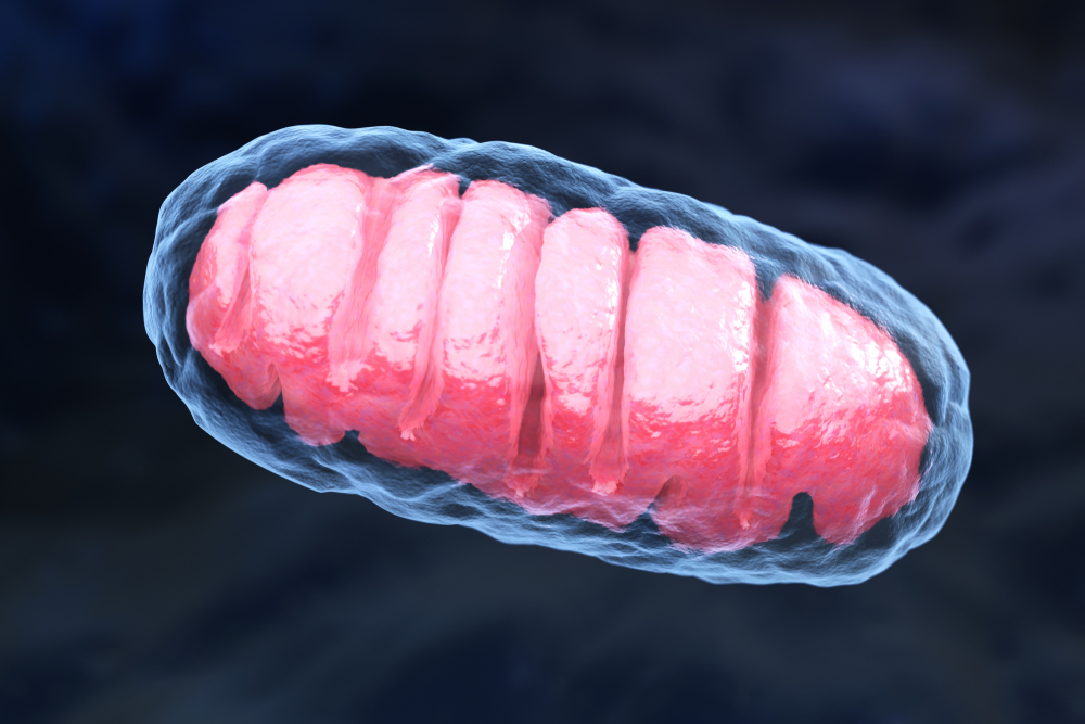
Researchers at Case Western Reserve University School of Medicine in Cleveland found that increasing levels of mitofusin 2 (Mfn2) — a protein that regulates the shape and structure of mitochondria, the cell compartments that produce energy — in the spinal cord of mice with ALS prevented muscle atrophy and paralysis.
“We found a way to alleviate age and ALS-related muscular atrophy in our mouse models,” Xinglong Wang, PhD, an associate professor of pathology, said in a press release. “Amazingly, we could delay ALS symptom onset by 67 days.”
First, investigators found that both older mice and those that were an ALS animal model — with symptoms similar to the human disease —had lower-than-usual levels of Mfn2 in their spinal cords.
Next, the researchers genetically engineered the nerve cells that connect the spinal cord to muscle fibers in the diseased and aged mice so these cells produced Mfn2 at unusually high levels. The increase prevented diseased animals from developing muscle atrophy, gait abnormalities, or losing evidence of grip strength — all symptoms that non-engineered control ALS mice acquired.
“Upregulation [higher production] of Mfn2 specifically in nerve cells is sufficient to abolish skeletal muscle loss in ALS and aged mice, despite ALS-causing protein being found in all organs and tissues,” Wang said.
They also found that Mfn2 helps to carry calpastatin — a nutrient that blocks harmful enzymes (a type of protein) from destroying nerves and muscle fibers. Mfn2 is found on the surface of mitochondria and travels along nerve cells extensions, called axons, to deliver nutrients to neuromuscular junctions. Without Mfn2, mitochondria fail to transport calpastatin to these points where nerve cells and muscle fibers “communicate,” leading to nerve damage and muscle atrophy.
“We found mitochondria function as miniature ‘trucks’ to transport protein along axons to prevent synaptic [nerve cell communication] degeneration,” Wang said.
Researchers suggested their work may have broad treatment implications — because problems with Mfn2 (low levels or evidence of mutations) are often found in ALS patients and those with other neuromuscular diseases or nerve damage. Restoring or even supplementing protein levels may be of clinical benefit.
“Our results suggest that, as a potential key component of a novel and heretofore unrecognized mechanism of cytoplasmic protein transport, Mfn2 may play a general role in preserving neuromuscular synapses and serve as a common therapeutic target for skeletal muscle atrophy,” the team concluded.