
Motor neurons’ size may make them vulnerable to ALS
Researchers create a cellular and gene expression atlas of the adult spinal cord
Written by |
Human motor neurons rely on a specialized molecular architecture to help support their large size, which may partially explain why they are more vulnerable than other nerve cells to damage in amyotrophic lateral sclerosis (ALS).
That’s according to the study, “A cellular taxonomy of the adult human spinal cord,” which was published in Neuron.
ALS is caused by the progressive death and degeneration of motor neurons, the specialized nerve cells that are responsible for controlling muscle movements. While numerous risk factors for ALS have been identified, it hasn’t been clear why motor neurons in particular are so vulnerable to damage in ALS.

A team of scientists supported by the National Institutes of Health (NIH) now conducted a study seeking to characterize the neurons and other types of cells inside of the human spinal cord, with a particular eye toward understanding why motor neurons are more vulnerable to ALS-related damage than other types of cells.
“As a resource, this cellular and gene expression atlas of the adult human spinal cord makes it possible to register known molecular [markers of disease processes] with human spinal cell types,” the team wrote.
Using post-mortem spinal cord samples from 14 people, the researchers conducted single-cell RNA sequencing. Simplistically, this method allows scientists to evaluate the overall gene expression profiles of individual cells, determining which genes are active and to which extent, and which genes are turned off.
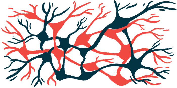
The study included data for more than 55,000 individual cells across the 14 samples. Based on patterns of gene expression, the team identified 35 populations of neurons — including one group of motor neurons and 34 other distinct types of nerve cells.
Compared with the other types of nerve cells, motor neurons showed higher levels of genes that have been linked with the development of ALS. Motor neurons also showed markedly higher levels of genes that are related to the cytoskeleton — the scaffold of proteins that helps to give cells their shape — and higher levels of neurofilament genes, which are needed to support cells growing to large size.
Motor neurons are large cells. Their axons (nerve fibers) can be up to a meter long, which is needed so they can stretch out to the muscles they control. These findings collectively imply that motor neurons rely on specialized molecular structures to support their large size, and that this may make them especially vulnerable to degeneration in diseases like ALS.
“Overall, these findings support a model of specific molecular repertoires for [motor] neuron cell structure that also confer selective vulnerability to degeneration,” the scientists concluded.
Clusters of support cells
In addition to the neuron populations, the team also identified 29 clusters of non-neuron support cells in the spinal cord. These included immune cells, blood vessel cells, and structural cells, as well as glia, a broad group of nervous system cells that help to support neuronal function.
“It is important to consider the spinal cord as a community of cell types that function together in normal health and disease,” the scientists wrote.
The team has made the comprehensive spinal cord cell atlas dataset available online for other researchers to use.
“We hope this work, together with other ongoing efforts, will serve as a foundation for studying the wide range of cell types involved in human spinal cord function,” they wrote.


Leave a comment
Fill in the required fields to post. Your email address will not be published.