
Magnetic stimulation seen to restore ALS motor neurons in cell study
Scientists say results may lead to potential novel therapy for ALS
Written by |
Magnetic stimulation restored the health of lab-grown motor neurons — the muscle-controlling nerve cells that die in amyotrophic lateral sclerosis (ALS) — derived from people with familial ALS, according to a proof-of-concept study.
The approach improved several biological processes in motor neurons, including the movement of cellular components along nerve fibers, or axons, and the growth of new axons.
“In the numerous series of experiments, we were able to demonstrate that the motor neurons from ALS patients respond to the magnetic fields,” Arun Pal, a cell biologist at the Helmholtz-Zentrum Dresden-Rossendorf (HZDR) research center, in Germany, who served as the study’s co-senior author, said in a university press release.
Thomas Herrmannsdörfer, PhD, another study author from the HZDR, noted that “detailed follow-up studies are required to corroborate [these] findings,” but added, “We regard these … results as an encouraging approach on the path to a potential novel therapy for ALS as well as other neurogenerative diseases.”
The study, “Restoring Axonal Organelle Motility and Regeneration in Cultured FUS-ALS Motoneurons through Magnetic Field Stimulation Suggests an Alternative Therapeutic Approach,” was published in the journal Cells.

Magnetic stimulation applied to lab-grown motor neurons
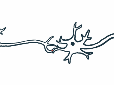
Motor neurons are nerve cells in the brain and spinal cord that send signals to muscles to perform various bodily functions, including movement, speaking, swallowing, and breathing. Their axons — the cable-like part that snakes away from the main part of the cell — are the longest in the body and can stretch from the spinal cord to the toes.
Most cases of ALS are characterized by the sporadic and progressive loss of motor neurons. In about 10% of cases, however, inherited genetic mutations are responsible for motor neuron death.
Magnetic stimulation is a non-invasive procedure that uses magnetic fields to stimulate nerve cells. It’s been approved for use in psychiatric disorders, such as depression.
A small clinical trial recently showed that magnetic stimulation slowed muscle function decline in ALS patients.
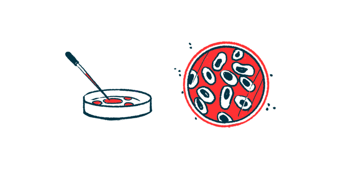
To investigate its impact at the cellular level, researchers in Germany now applied magnetic stimulation directly to lab-grown motor neurons derived from three ALS patients carrying inherited disease-causing mutations in the FUS gene.
Motor neurons were grown from patient-derived induced pluripotent stem cells, called iPSCs. Such iPSCs are generated from fully matured cells that are reprogrammed back to a stem cell-like state, where they can give rise to almost every type of human cell.
The team previously showed the movement of cellular components, or organelles, along axons was impaired in spinal motoneurons derived from FUS-ALS patients. These organelles included mitochondria, the cell’s energy producers, and lysosomes, the cell’s recycling centers.
Now the researchers found that magnetic stimulation restored the movement of these organelles in lab-grown patient-derived motor neurons without affecting cells derived from three healthy people. Restoration depended on the frequency of the magnetic field, with a combination of 2 and 10 Hz (cycles per second) generating the best results.
“The axonal transport of mitochondria (the power stations of the cell) and other organelles that are impaired in ALS cells is reactivated by stimulation with magnetic fields,” Pal said.

Scientists now planning clinical pilot studies
In FUS-ALS mouse models, the growth, branching, and distribution of axons — which go on to form connections to other nerve cells as well as muscle cells — have been shown to be impaired.
Consistently, untreated patient-derived motor neurons showed slower outgrowth than those derived from healthy controls.
With the 10 Hz treatment, axons of patient-derived motor neurons grew as fast as control cells, while the 2 Hz and 2/10 Hz combination did not show significant changes. No evidence of magnetic stimulation-related adverse effects on patient and control motor neurons was observed.
These findings showed that, with magnetic stimulation treatment, “axonal regeneration — which is the ability to regrow and reconnect — can be restored,” Pal said.
The shape of nerve cells is unique, with axons extending from the cell body. This shape is dictated by the cytoskeleton, a complex network of interlinking proteins called microtubules that helps cells maintain their shape and internal organization.
The levels of a microtubule stabilization marker were significantly lower in FUS-ALS motor neurons relative to controls. With magnetic stimulation, marker levels in patient cells rose back to control levels, pointing to “an improvement of microtubule stability that is feasibly beneficial for deficient axonal organelle trafficking and regeneration,” the researchers wrote.
“This work provides proof of concept demonstrating that magnetic field treatments are powerful in restoring several hallmarks of ALS [development] recapitulated in our cultured disease model of human iPSC-derived spinal [motor neurons],” the team concluded.
“Long-term and [living organism] studies are required to further substantiate the therapeutic potential of magnetic field treatments,” they added.
The scientists are now planning clinical pilot studies to assess the therapeutic potential of magnetic simulation, applied with specialized equipment.

Leave a comment
Fill in the required fields to post. Your email address will not be published.