
Muscle Imaging Helps Evaluate Nerve Loss in ALS and Other Motor Diseases, U.K. Study Finds
Written by |
Analyzing muscles via magnetic resonance imaging (MRI) can help detect small physiological changes that happen when motor neuron diseases like amyotrophic lateral sclerosis (ALS) get worse, concludes a study by British researchers.
The study, “Imaging muscle as a potential biomarker of denervation in motor neuron disease” appeared in the Journal of Neurology, Neurosurgery & Psychiatry.
Understanding clearly how a disease progresses, and finding accurate measures of disease progression, are two key aspects to developing new and effective therapies. But in the field of motor neuron diseases, objective measures of such disease progression are still lacking.
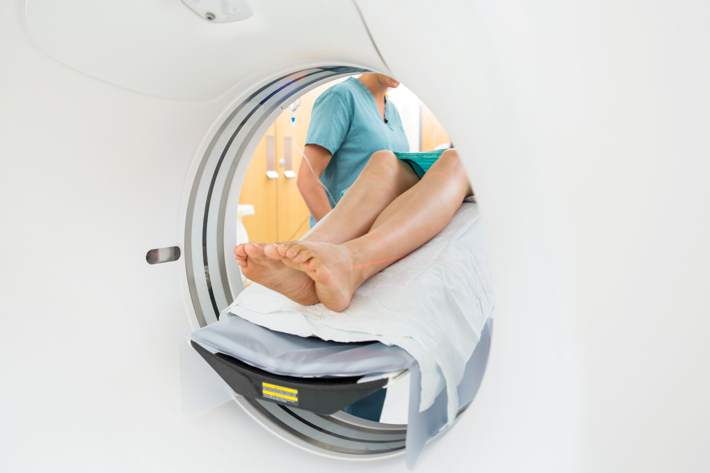
In these diseases, patients lose the nerve cells, called motor neurons, that deliver brain signals to muscles. This is accompanied by fluid shifts, independent of a patient’s physical efforts, that can be detected with MRI imaging exams.
A team of researchers at England’s University of Sheffield investigated if MRI imaging of whole-body muscles could accurately detect small progressive changes in patients with motor neuron disease.
The study recruited 26 ALS patients, three patients with progressive muscular atrophy and 22 healthy volunteers. Participants underwent MRI analysis and standard muscle assessment methods at the start of the study, and then four to six months after enrolment.
At baseline, patients already had less muscle potential and fewer nerve cells in the biceps and lower leg muscle than did control individuals. MRI evaluation also showed that patients had increased muscle denervation – when the connection between nerve and muscle cells is lost.
Consistently, increased MRI signals indicative of muscle denervation were associated with greater muscle weakness and lower muscle potential. interestingly, MRI scores were associated with progressive disease in the legs but not in biceps.
In the follow-up analysis, the control group had no significant changes in MRI or muscle function scores. But researchers observed increased MRI scores and denervation index in motor neuron disease patients and decreased lower-leg muscle power.
Overall, researchers believe that MRI evaluation of lower-leg muscles reflects clinically relevant aspects in the progression of motor neuron diseases, and that it can be used as an objective measure of disease progression.
“Whole-body muscle MRI offers a new approach to objective assessment of denervation over short timescales in MND [motor neuron disease] and enables investigation of patterns of disease spread in vivo,” researchers concluded.


Charlie
This tells us nothing new that would help.
It is simply 'symptom identification.'
News that ALS progression/decline correlates to decreasing muscle ability is what pALS experience every day.
Hey Sheffield, tell us something we don't know.
sunita joshee
is it helps in treatment of mnd if yes plz share us how it is possible and what is treatment and from where we can approch to reach
Hannan Ahmad
Just like as MRI has been helping in the treatment of knee injuries and bone density problems, this is a new dimension about how this amazing technology will get implemented.
elke jaquet
i have all the symptons of MND, but neither an MRI, nor lumber puncture confirmed that i suffer from MND