ALS Affects More Brain Regions Than Just the Movement Control Area, Study Shows

Scientists have long believed that ALS was confined to one area of the brain, but an Irish study shows it affects a number of regions.
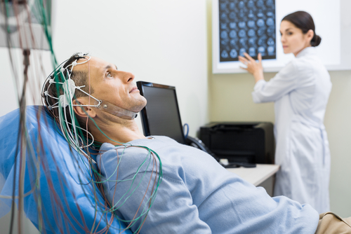
Researchers at Trinity College Dublin’s Academic Unit of Neurology used electroencephalography, or EEG, to record patients’ brain activity. They discovered that nerve cell communication was lower than normal in some regions and higher in others.
The study, “Characteristic Increases in EEG Connectivity Correlate With Changes of Structural MRI in Amyotrophic Lateral Sclerosis,” was published in the journal Cerebral Cortex.
ALS is a neurodegenerative disease that mainly affects muscle movement, like chewing, walking, breathing and talking. It gets worse over time, and is fatal.
While scientists long considered it a disease that affected only the brain’s movement-control region, recent evidence has suggested that it affects non-movement-related areas as well.
Irish researchers decided to see exactly which brain areas that ALS affects. Their study involved 100 ALS patients and 34 healthy volunteers.
Using electroencephalography to measure the brain’s electrical waves has helped scientists understand conditions like epilepsy. But while it is inexpensive and easy to use, researchers have not used it a lot to study progressive neurologic diseases like ALS.
“Understanding how the networks in the human brain interact in health and disease is a very important area that has not been adequately researched,” Dr. Bahman Nasseroleslami, a senior research fellow at Trinity who was lead author of the study, said in a news release.
ALS patients’ EEG patterns showed changes in nerve cell connectivity, researchers said. The patterns persisted over time, suggesting they were related to the disease’s progression.
The team also compared the EEG results with brain structure data they obtained with magnetic resonance imaging (MRI) scans of patients’ brains.
The brain regions where EEGs uncovered loss of communication activity were the same ones where MRIs spotted ALS-related structural degeneration. In contrast, areas with less degeneration had better nerve cell communication, the team said.
“Using EEG to decipher changes in brain function has not been possible until recently,” Nasseroleslami said. “The computational power, mathematical and statistical tools were just not available.”
But advances in technology and mathematical analysis mean that “we can now explore the living human brain in a very sophisticated and non-invasive way, and that we can link our dynamic EEG changes with anatomical changes captured by MRI.”
“This expands enormously our ability to understand how the brain is working in real time, and how these changes in brain networking correlate with structural changes that we can see on MRI scans,” he added.
The study’s key takeaway is that ALS is similar to other neurodegenerative diseases in that its nerve cell connectivity changes appear in several brain regions rather than just one.
“Our findings will revolutionize how we measure changes in brain function in ALS and many other related neuro-degenerations such as frontotemporal dementia,” said Professor Oria Hardiman, who heads Trinity’s neurology program. “Our findings will also help in understanding the links we have shown previously between ALS and schizophrenia.
“There is much to do, but this is the first step in developing new and innovative measurements that will have a major impact on how we conduct future clinical trials” of potential therapies, he added.


