
Cholesterol Transporter May Be Strongest Trigger of Sporadic ALS
ApoB100 protein in cerebrospinal fluid of sALS patients promoted disease in mice
Written by |

The cerebrospinal fluid of people with sporadic amyotrophic lateral sclerosis (sALS), but not of those with familial forms of the disease, promoted ALS-specific molecular, cellular, and motor abnormalities in healthy mice, a study showed.
Apolipoprotein B-100 (apoB100), a protein involved in cholesterol transport, was identified as the main factor responsible for these damaging effects. A single administration of apoB100 was sufficient to recapitulate ALS’s cellular and clinical hallmarks, including motor disability and motor neuron loss, while removing apoB100 from the cerebrospinal fluid of sALS patients lessened its neurotoxic effects.
The cerebrospinal fluid, or CSF, is the liquid that surrounds the brain and spinal cord.
“The impact of this research is potentially groundbreaking for patients with sporadic ALS, the predominant form of this devastating disease,” Saud A. Sadiq, MD, the study’s senior author at the Larry G. Gluck division of ALS research, part of the Tisch Multiple Sclerosis Research Center of New York, said in a center press release.

“For the first time ever, our novel animal model has identified the protein responsible for causing debilitating symptoms like motor disability and motor neuron degeneration in sALS,” added Sadiq, who is also Tisch’s director and chief research scientist.
Findings, he added, suggest that “targeted reduction of this protein from [the] CSF could open up a new and potentially promising avenue to treat thousands of ALS patients.”
The study, “Apolipoprotein B-100-mediated motor neuron degeneration in sporadic amyotrophic lateral sclerosis,” was published in the journal Brain Communications.
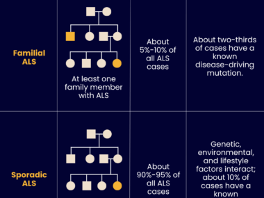
ALS is caused by the progressive loss of motor neurons, the specialized nerve cells that control voluntary movement. Both genetic and environmental factors are thought to contribute to its development, with only about 10% of all cases being considered familial (fALS), due to inherited disease-causing mutations.
While several molecular and cellular abnormalities have been implicated in ALS, whether they have the same degree of importance in sporadic and familial forms is unclear, and therapeutic options for its most common form are lacking.
CSF of sporadic ALS seen to differ in effects from that of familial ALS
Previous studies showed that injecting cerebrospinal fluid from ALS patients into mice could promote ALS-like disease. But the specific CSF components responsible for these neurotoxic effects remains unknown. It also is unclear whether related differences exist in the CSF of sALS and fALS patients.
Sadiq and his team, along with Merit E. Cudkowicz, MD, compared the neurotoxic effects of CSF taken from 11 sporadic ALS patients and seven people with familial ALS in a newly created CSF-mediated mouse model.
Cudkowicz is the director of the Sean M. Healey and AMG Center for ALS at Massachusetts General Hospital (MGH) in Boston, and the principal investigator of the HEALEY ALS platform trial (NCT04297683) that is testing several ALS treatment candidates simultaneously. Twelve of the study’s patients were seen at this center, whereas the remaining six were followed at the Gluck center.
Patients with familial ALS carried mutations in one of three disease-associated genes: SOD1 (three patients), C9orf72 (three patients), and TARDBP (one patient).
A single injection of patients’ CSF was administered directly into the spinal canal of healthy adult mice, and disease-associated molecular, cellular, and motor function changes were assessed.
Results showed that CSF from sALS patients, but not from those with familial forms, promoted rapid and permanent motor disability, all associated with hallmark features of ALS.
These included motor neuron loss, abnormal localization of the TDP-43 protein, and abnormal activity of brain cells that included astrocytes and microglia, both types of glial cells essential for a healthy central nervous system.
Notably, some of these molecular and cellular abnormalities were also observed in mice injected with CSF from patients with C9orf72 and/or those with TARDBP mutations, but these were considerably milder.
“The moderate extent of neurotoxic effects induced by C9orf72 and TARDBP CSF, as well as the lack of effects induced by SOD1 CSF confirms the specificity of our sALS animal model,” the researchers wrote.
ApoB100 likely at ‘neurotoxic levels’ in sporadic ALS patients
To identify potential neurotoxic candidates in the fluid of sALS patients, the team systematically filtered out its components by size and reassessed the effects of the filtered CSF in the mouse model.
This, combined with a comprehensive analysis of all the proteins in the CSF pre- and post-filtration, allowed the identification of ApoB100 “as the neurotoxic protein responsible for inducing motor disability and motor neuron degeneration,” the team wrote.
Apolipoprotein B-100 is a component of the particles responsible for transporting cholesterol and other fatty molecules in the blood.
Importantly, injecting human ApoB100 alone in the spinal canal of healthy mice was sufficient to recapitulate the disease-specific molecular, cellular, and clinical abnormalities seen with sALS CSF. Exposure to this protein was also found to promote death in lab-grown human motor neurons.
Similar experiments using other proteins identified in the screenings or those previously implicated in neurodegenerative conditions did not result in ALS-like features.
Targeted removal of ApoB100 from sALS cerebrospinal fluid, either via filtration or the use of specific antibodies, prevented motor neuron loss and the development of motor deficits in the mouse model.
However, other ALS features, such as abnormal activity of astrocytes and microglia, were comparable between mice given unchanged and ApoB100-depleted sALS CSF. This suggested that components other than ApoB100 contribute to the disease.
“This study presents apolipoprotein B-100 as a novel therapeutic target specific for the predominant sporadic form of amyotrophic lateral sclerosis,” the researchers wrote. It establishes “proof-of-concept to support targeted reduction of ApoB via CSF [filtration] as a therapeutic strategy for sALS patients.”
Findings also highlight “fundamental differences” in the underlying mechanisms of sporadic and familial ALS, “with genetic mutations perhaps increasing intrinsic vulnerability of motor neurons to stress in fALS, while neurotoxic levels of ApoB circulating in CSF contributes to neurological damage in sALS,” the scientists wrote.
“This emphasizes the necessity of tailoring therapeutic approaches to specific ALS subtypes and the importance of resolving the toxic CSF environment in sALS,” they added.
Jamie K. Wong, PhD, a research scientist at Tisch and the study’s lead author, said: “We are excited to build upon the foundation established with this novel sALS animal model, as we now have the opportunity to further investigate sALS CSF-induced [disease] mechanisms and test potential therapies in a sALS-specific model.”
These scientists are now using this type of mouse model to study multiple sclerosis, another neurodegenerative disease, and Wong believes “this approach may potentially be useful for studying other neurodegenerative diseases as well.”


Leave a comment
Fill in the required fields to post. Your email address will not be published.