
This immune protein plays key role in nerve damage, early study finds
Suppressing the protein GSDME prevented nerve fiber loss in patient-derived cells
Written by |

An immune protein that is mostly produced in neurons was found to drive damage to mitochondria, which produce energy for cells, and nerve fiber loss, a study has found.
Suppressing the protein, called gasdermin-E (GSDME), prevented nerve fiber loss in cells derived from people with amyotrophic lateral sclerosis (ALS), and also prolonged survival and improved motor function in an ALS mouse model.
Researchers noted that understanding the details of GSDME-mediated nerve damage may support the development of new therapeutic strategies for neurodegenerative diseases.
“The unmet need for therapies for neurodegenerative diseases is huge, and our work opens up a whole new pathology that we could address,” Judy Lieberman, MD, PhD, professor at Harvard Medical School (HMS) and study co-lead said in a university press release.
“We describe a pathway and molecules that you can target for treating many neurodegenerative diseases,” said Lieberman, who is also chair of cellular and molecular medicine at Boston Children’s Hospital.

The discovery was described in the study “Gasdermin-E mediates mitochondrial damage in axons and neurodegeneration,” which was published in the journal Neuron.
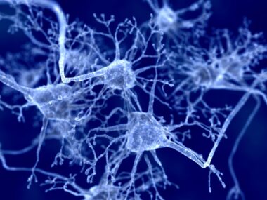
ALS is marked by the shrinkage and death of motor neurons, the nerve cells that send messages between the nervous system and muscles to control voluntary movements. In neurological diseases such as ALS, nerve cell death is often preceded by the impaired function of mitochondria and the loss of nerve fibers called axons.
Gasdermins are proteins linked to cell death
Gasdermins (GSDMs) are a family of proteins linked to cell death via a process called pyroptosis — a form of death triggered by the formation of pores in the cell membrane. Pyroptosis occurs most often upon infection and is thought to be part of the antimicrobial response, but it has also been associated with disease-related inflammation.
“Inflammation is a double-edged sword and could be either protective or destructive based on context,” said Isaac Chiu, PhD, associate professor of immunology at HMS and study co-lead.
GSDME is a GSDM family member produced in the brain and spinal cord, but its role in nerve cell function remains largely unknown. The team at Harvard set out to address that question.
The protein was found to be activated when neurons were exposed to chemicals that are toxic to mitochondria to model features of neurodegeneration. GSDME specifically targeted the membranes within mitochondria, where it caused pores that released the mitochondrial content to mediate neuronal damage.
Consistently, mouse neurons modified to lack GSDME showed greater mitochondrial integrity and resistance to the toxic chemicals. Overall, evidence indicated that GSDME regulated the integrity of axons, and when activated locally, it destroyed mitochondria and damaged axons without affecting neuron cell bodies.
“If you look at a plate of neurons, you see a jungle of axons,” said Himanish Basu, PhD, co-first author of the study. “But if you look at a plate where gasdermin E is activated, you see retractions of these cellular processes.”

GSDME contributed to mitochondrial dysfunction, axon loss
To understand whether GSDME could contribute to the neuronal death caused by ALS-associated proteins, the team grew mouse and human-derived neurons with TDP43 and PR-50 — a product of C9ORF72 mutations. This exposure induced GSDME activation and caused mitochondrial dysfunction and axon loss.
In contrast, exposure to these proteins did not impact axons in cells lacking GSDME. The team noted these results mirrored those obtained using mitochondrial toxins.
“Our work is an example of how immunology can help explain neurodegeneration on a mechanistic level, and what drives axon loss and neuronal injury,” said Dylan Neel, a graduate student in Chiu’s lab and a co-first author.
Finally, the team tested the impact of GSDME in a standard ALS mouse model, which developed progressive loss of motor neurons in the spinal cord. The researchers detected activated GSDME in the spinal cords of symptomatic ALS mice, increasing further by the late stages of the disease, but the protein was not found in pre-symptomatic animals.
Deleting GSDME in ALS mice improved motor function, prevented axon and motor neuron loss, and reduced nerve-related inflammation, which ultimately extended the animals’ survival.
“We identify GSDME as an executioner of neuronal mitochondrial dysfunction that may contribute to neurodegeneration,” the researchers wrote. “Future work should determine the feasibility of targeting GSDME expression or function.”
Chiu added: “We revealed an innate immune molecule playing a role in neurodegeneration, which presents a new avenue of thinking about neuronal health.”
Leave a comment
Fill in the required fields to post. Your email address will not be published.